Published:
A clinical case from the practice of Professor Yakiv Vitaliiovych Fishchenko
Hanna Pavlivna, aged 87, sought treatment for longstanding lower back pain, pain in both legs and severely restricted walking. Before surgery, she could walk only 5–10 metres with support and had not left her home for more than six months. On 9 October 2026, Professor Yakiv Fishchenko performed biportal endoscopic decompression at L2–L3, L3–L4 and L4–L5 without stabilisation. At one month, her pain had decreased considerably and she could walk 100 metres with a cane.
Symptoms and limitations before surgery
The patient had experienced lower back pain and pain in both legs for many years, with symptoms worsening during walking. She also felt discomfort when lying down. Over the preceding year, her walking distance had decreased from approximately 100 metres to 5–10 metres with support. She had remained at home for more than six months.
Before surgery, lower back pain and pain in each leg were rated at 7–8 out of 10 on the visual analogue scale for pain (VAS). Her Oswestry Disability Index (ODI) was 82%. Her body mass index (BMI) was 29.1 kg/m².
Diagnosis and treatment planning
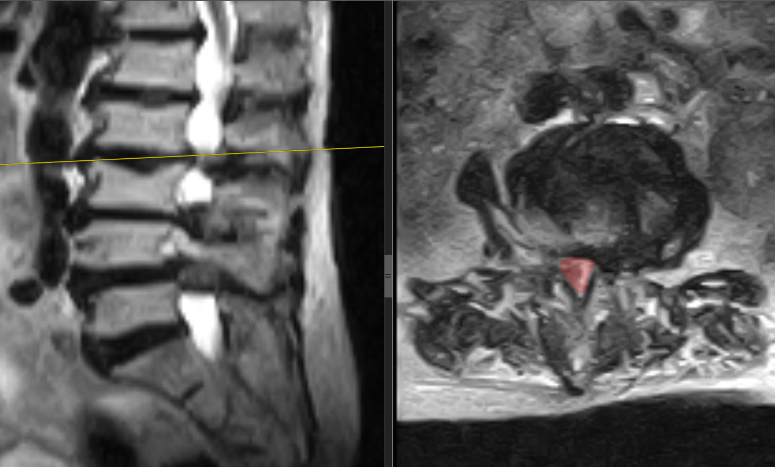
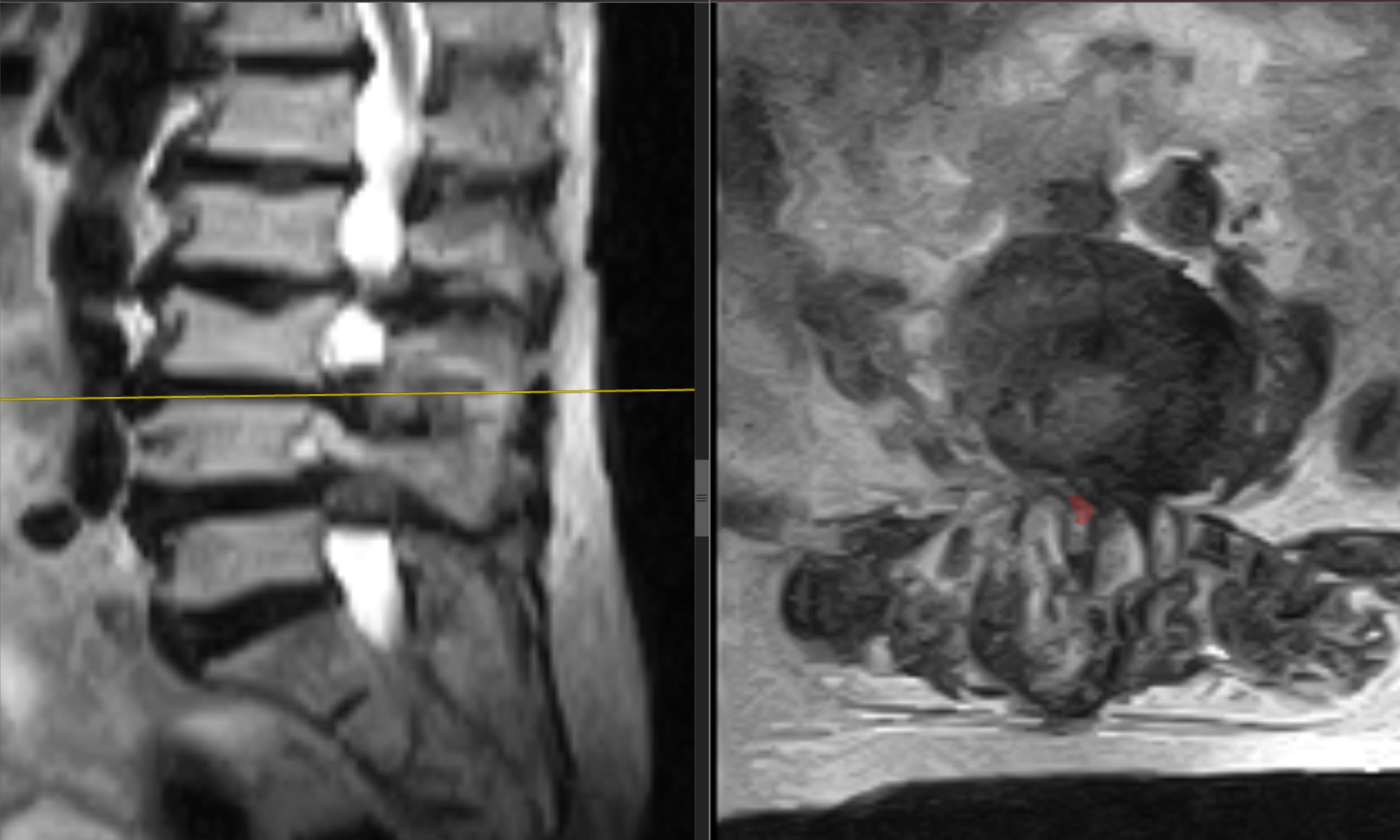
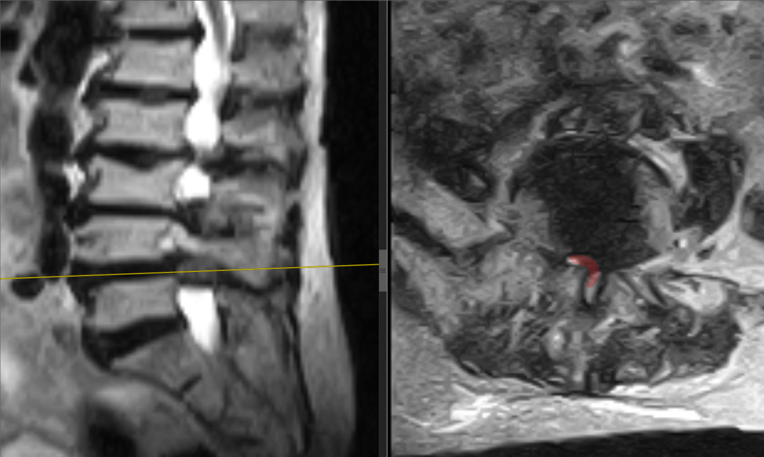
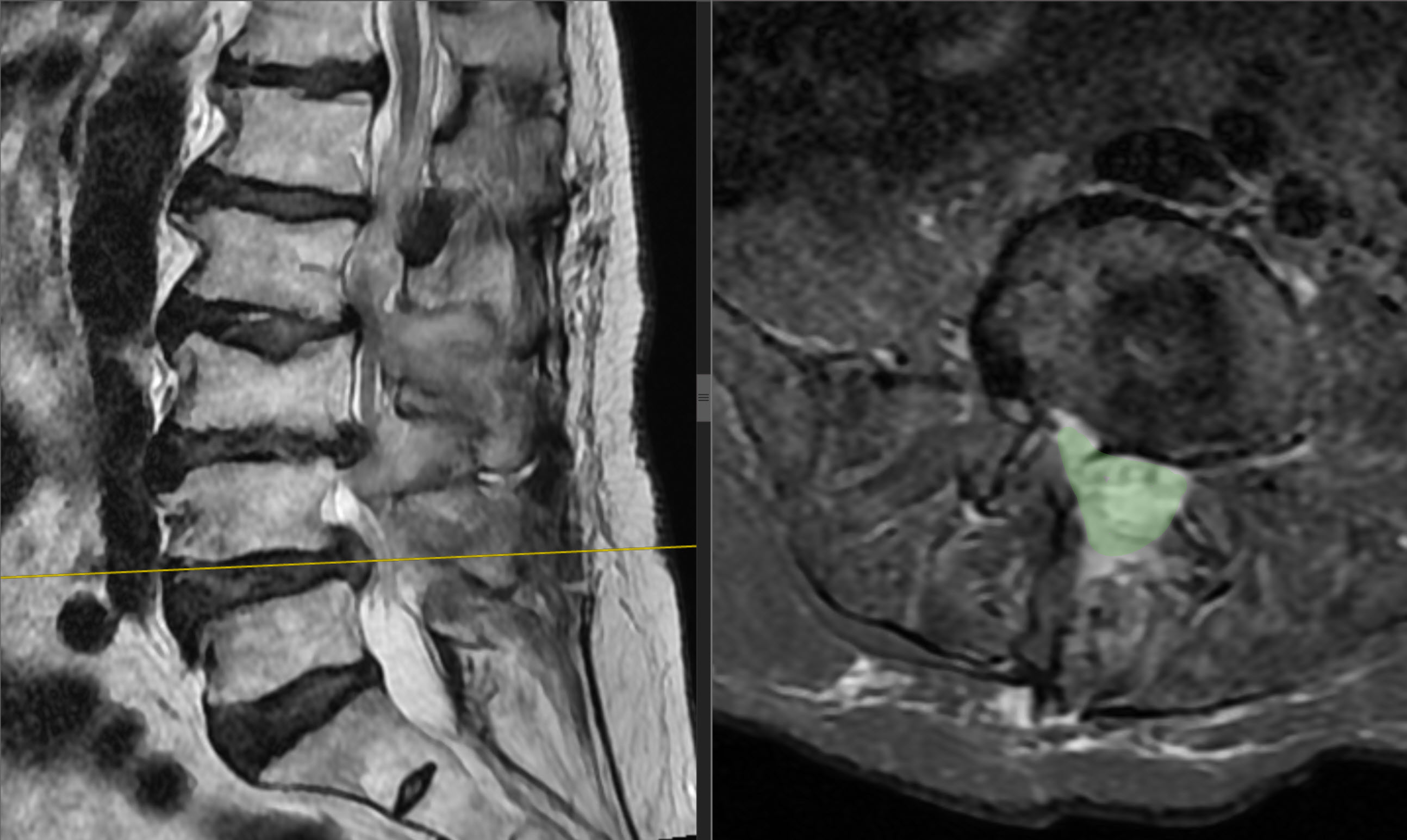
Lumbar spinal stenosis was diagnosed at three levels: L2–L3, L3–L4 and L4–L5. Mild degenerative scoliosis was present alongside marked degenerative changes in the intervertebral discs. No signs of instability were identified at the levels to be operated on. The patient also had marked osteoporosis.
Why decompression without stabilisation was chosen
Surgical planning took into account the absence of instability at these levels, the patient's age and marked osteoporosis. In the surgeon's assessment, stabilisation in this situation would have introduced additional risks and could have made rehabilitation considerably more difficult. Endoscopic decompression without implants was therefore chosen. This was an individual decision about the extent of surgery for this patient.
Surgical treatment
On 9 October 2026, Professor Yakiv Fishchenko performed BESS/UBE biportal endoscopic decompression at L2–L3, L3–L4 and L4–L5. All three levels were treated in a single procedure. The total operating time was 92 minutes. No stabilisation or implant placement was performed. There were no intraoperative complications.
Early recovery after surgery
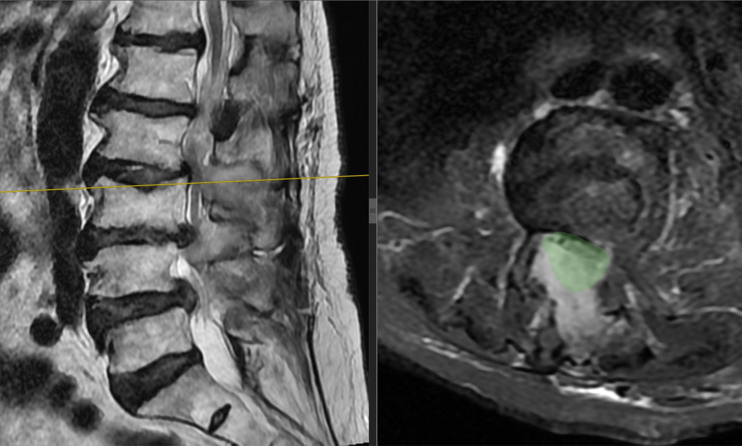
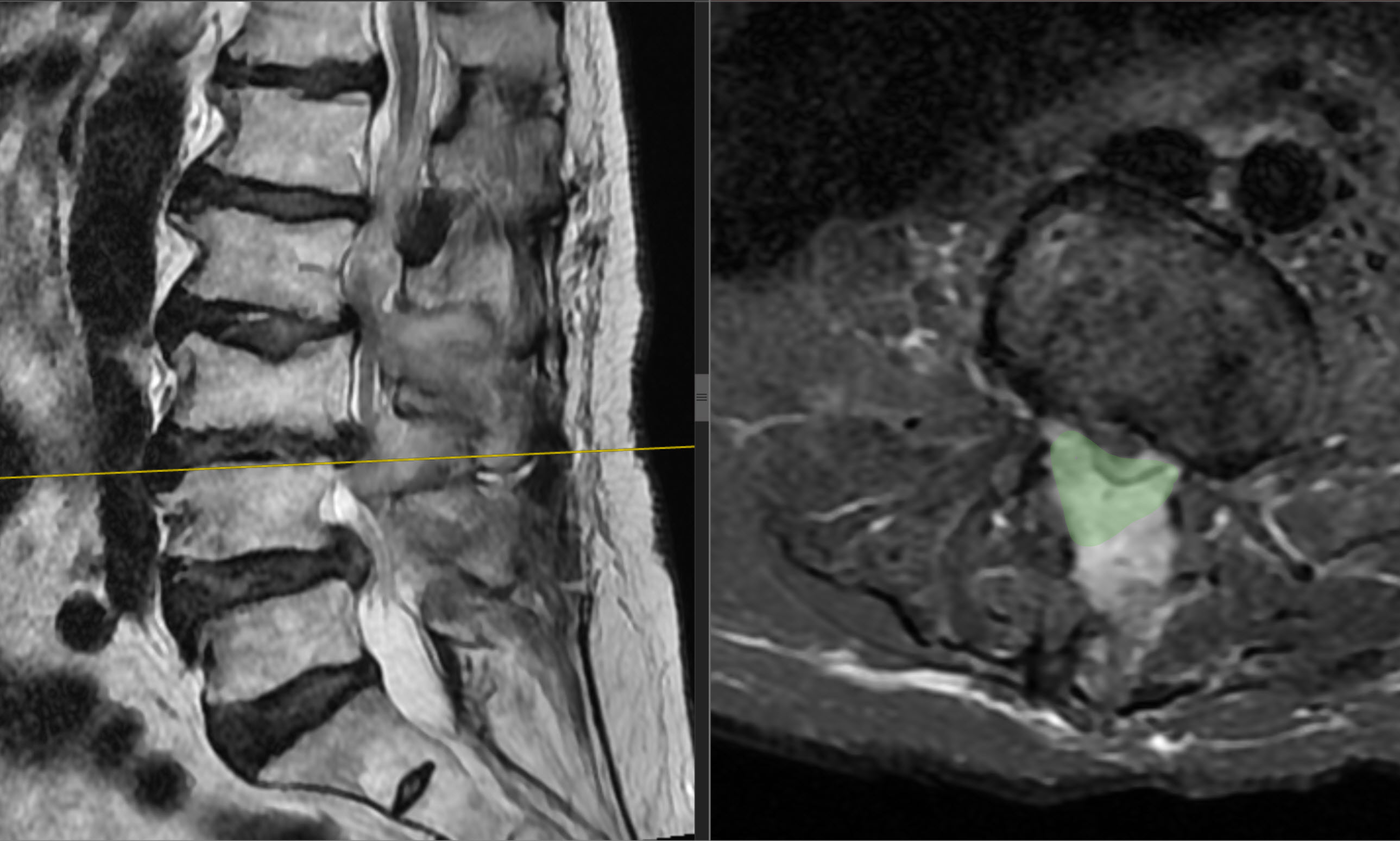
The patient got up and began walking the following day. A follow-up MRI was performed on 10 October 2026, the day after surgery. The early postoperative period proceeded as planned, without complications. She was discharged home on the second postoperative day.
Results at one month
At one month, lower back pain had decreased to 2/10, right leg pain to 2–3/10 and left leg pain to 1–2/10 on the VAS. ODI decreased from 82% to 36%, a reduction of 46 percentage points. Walking distance increased to 100 metres with a cane.
| Measure | Before surgery | At one month |
|---|---|---|
| Lower back pain on the VAS | 7–8/10 | 2/10 |
| Right leg pain on the VAS | 7–8/10 | 2–3/10 |
| Left leg pain on the VAS | 7–8/10 | 1–2/10 |
| ODI | 82% | 36% |
| Walking distance | 5–10 m with support | 100 m with a cane |
The one-month follow-up assessment documented a considerable reduction in pain and improved walking. The patient continued to use a cane; these findings do not indicate complete recovery or the absence of residual symptoms. They describe the outcome of this individual clinical case over one month of follow-up.
The patient's consent to publication of this clinical case and MRI images was obtained.
Clinical case discussion by Professor Yakiv Vitaliiovych Fishchenko.
MRI before and after surgery
The three MRI pairs below show preoperative images and follow-up scans from 10 October 2026. Within each image, the sagittal view is on the left and the axial view is on the right. The coloured markings were added by the author of the clinical case discussion.
Click an image to open it at full size.
More about treatment
More about lumbar stenosis treatment
Send MRI scans via Telegram for a consultation about surgical treatment
Discuss your case with the professor
Send a description of symptoms and existing investigations. If MRI/CT has already been performed, include the complete DICOM study and report. Preliminary review by the professor is free; an in-person consultation is also possible without an MRI already available.