Ten clinical cases from Professor Iakiv Fishchenko's practice: disc herniations, revision procedures, instability and lumbar stenosis. Separate pages provide clinical descriptions, investigations and results at the stated follow-up times.
Clinical cases: from investigations to follow-up
CASE 1
Right-sided L5–S1 herniation: a 73-year-old woman
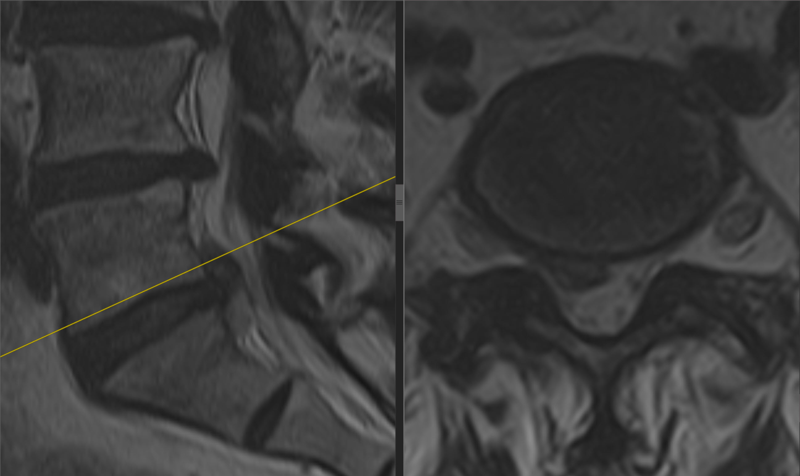
Biportal endoscopic discectomy through an interlaminar approach. Reported follow-up: one week.
Full discussion, images and video →CASE 2
Right-sided L5–S1 herniation: a 19-year-old man
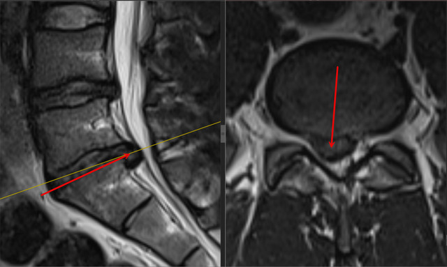
Right leg pain and foot weakness. BESS/UBE discectomy and one-week follow-up.
Full discussion, images and video →CASE 3
L5–S1 herniation with motor deficit
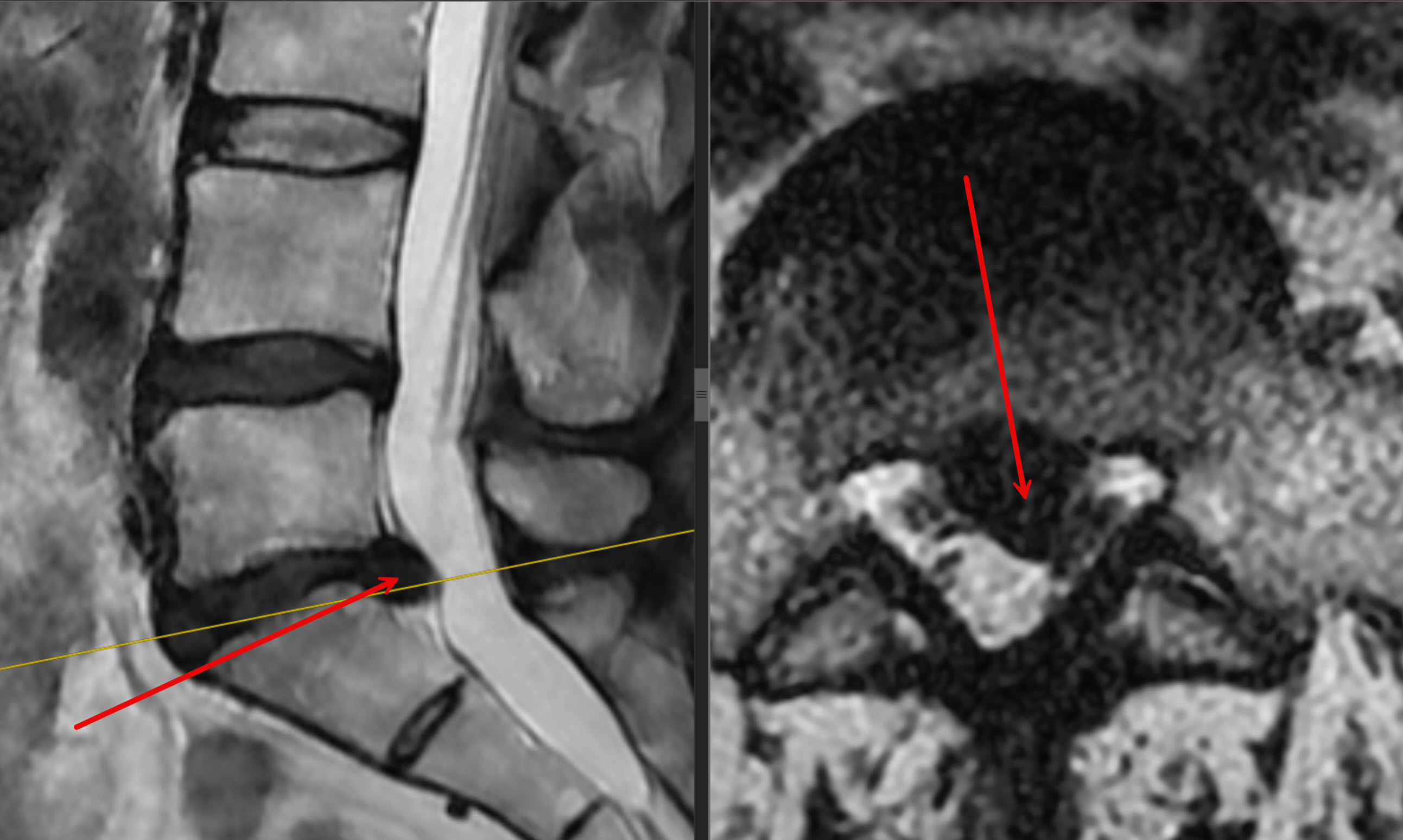
A 64-year-old woman: central L5–S1 herniation with left-sided symptoms, motor deficit and urinary dysfunction. Full updated discussion and one-week outcome.
Full discussion, images and video →CASE 4
Extraforaminal L4–L5 herniation
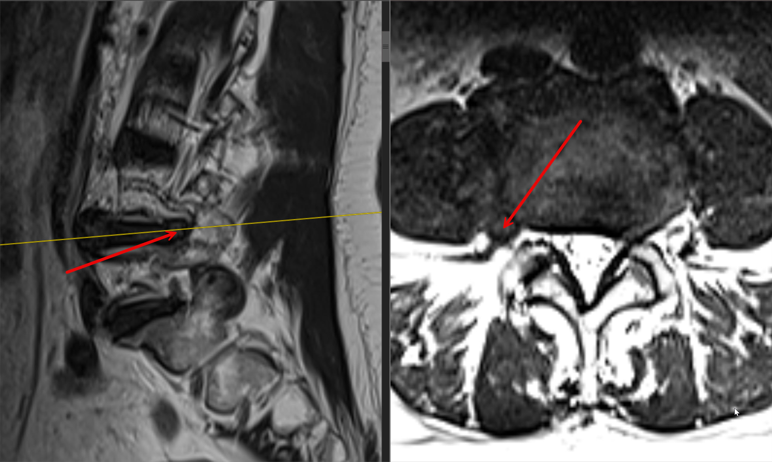
A 72-year-old woman with L4 radiculopathy. Transforaminal endoscopic discectomy and progress with neuropathic symptoms at one week.
Full discussion, images and video →CASE 5
L4–L5 instability: cage and fixation
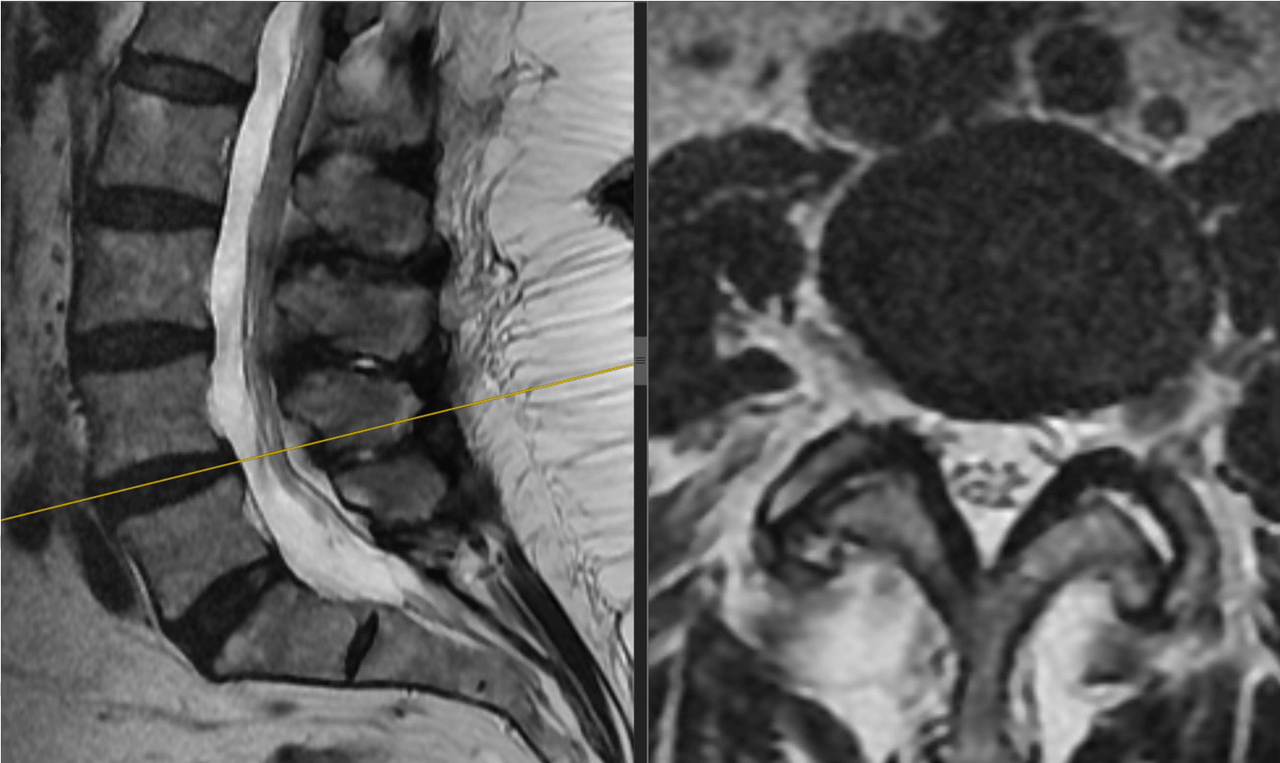
A 63-year-old woman. Endoscopic cage placement and percutaneous fixation without decompression. MRI, flexion–extension X-rays and animated CT.
Full discussion, images and video →CASE 6
L4–L5 stenosis: decompression without stabilisation
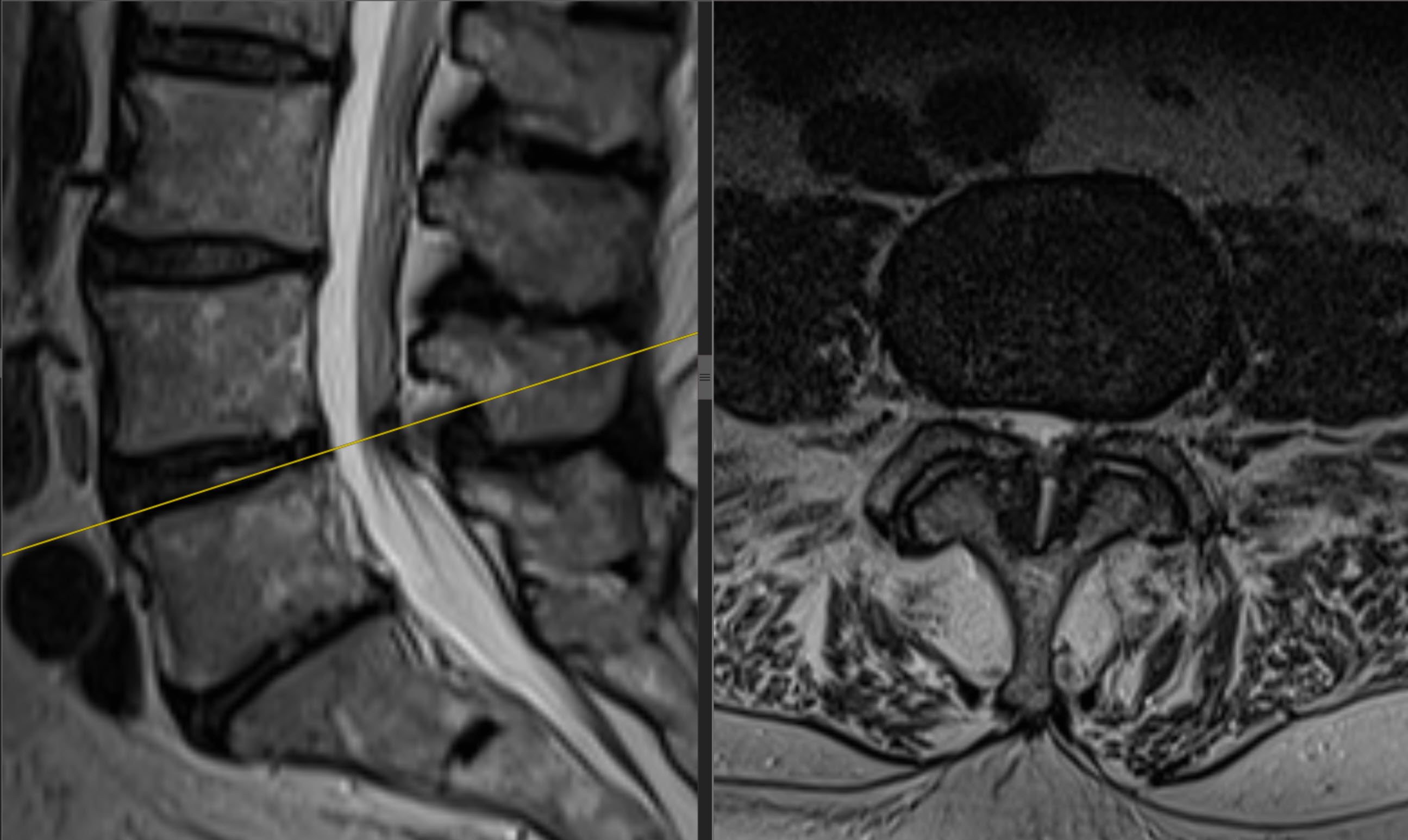
A 79-year-old woman. BESS/UBE decompression without fixation, pre- and postoperative MRI, and one-month follow-up.
Full discussion, images and video →CLINICAL CASE
L3–L4 stenosis: a 72-year-old man
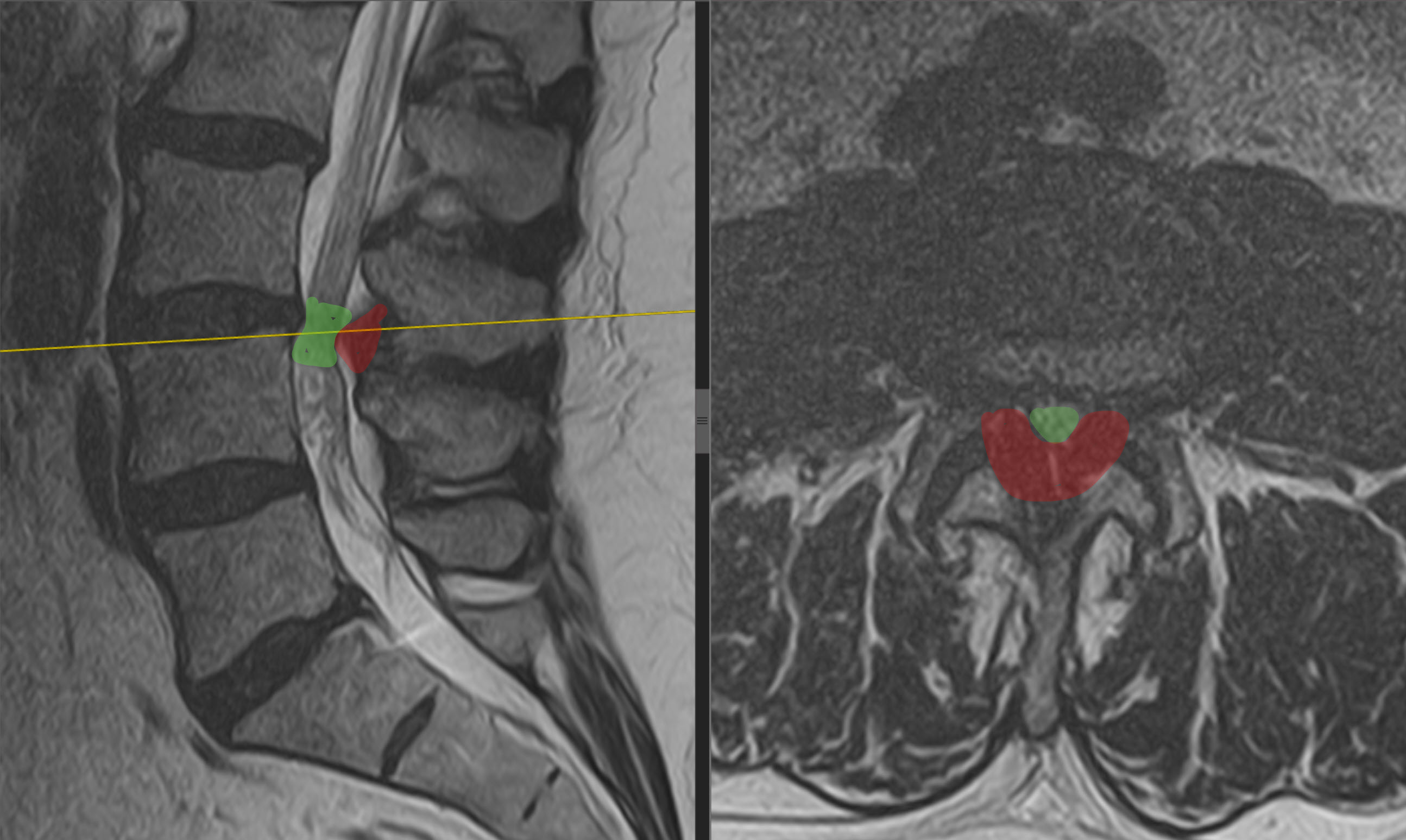
Walking limited to 200 metres before surgery. Biportal endoscopic decompression and results over the first two days.
Full discussion and MRI →CLINICAL CASE
Recurrent L4–L5 herniation: discectomy and L4–S1 stabilisation
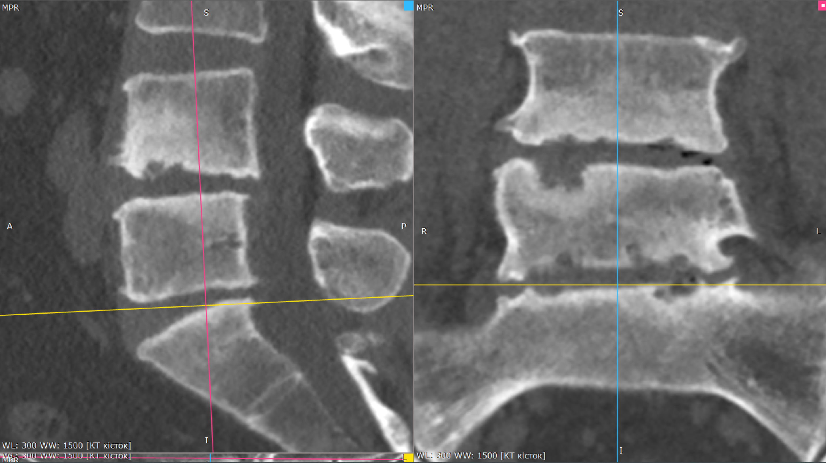
Revision endoscopic surgery with cages and fixation after symptoms returned. CT, MRI, X-rays and the early result on day seven.
Full discussion and investigations →CLINICAL CASE
Three-level L2–L5 stenosis: an 80-year-old woman
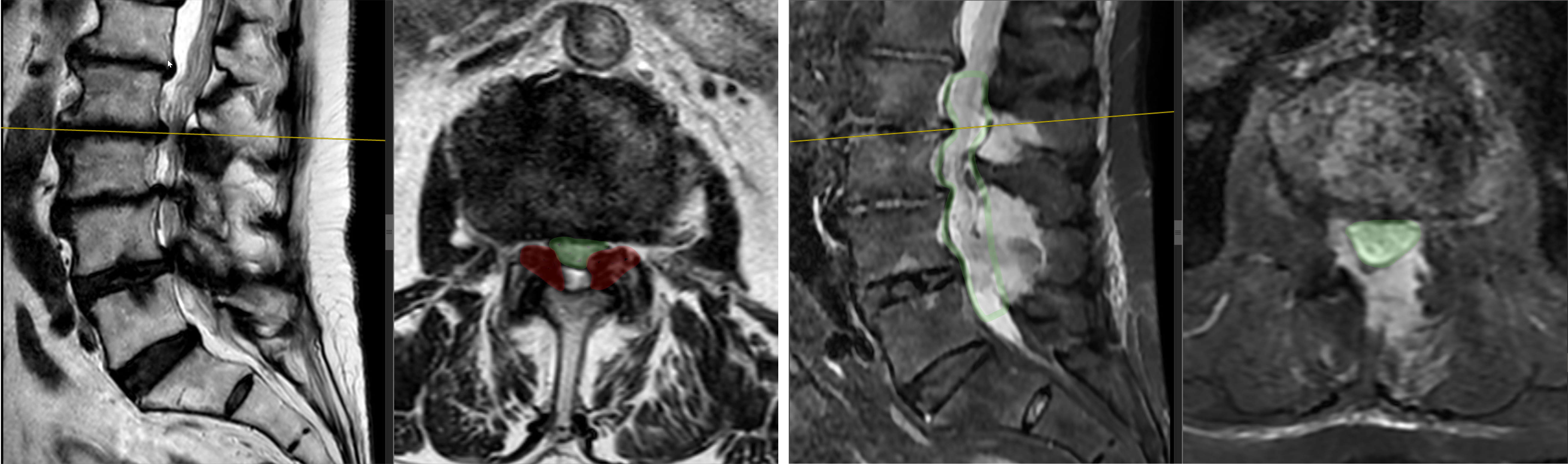
After endoscopic decompression, walking distance at one month was 150 m, compared with 10–15 m before surgery. Three MRI pairs and a table tracking pain, ODI and walking.
Full discussion and MRI →CLINICAL CASE
Three-level lumbar stenosis in an 87-year-old woman
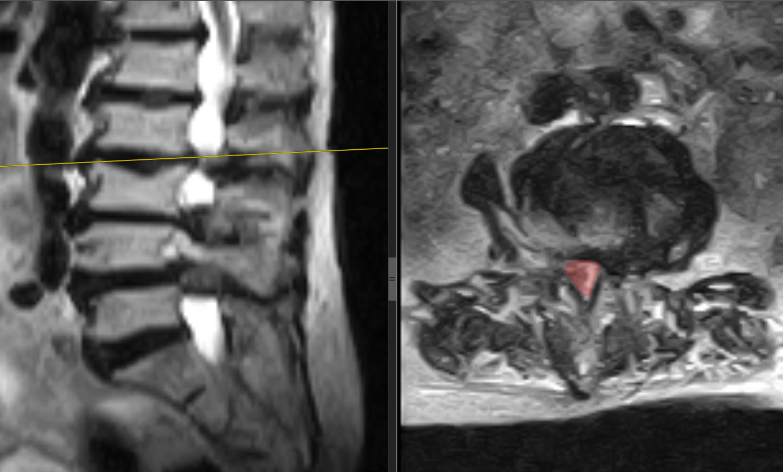
Three-level BESS/UBE decompression without implants. At one month, walking distance increased from 5–10 m with support to 100 m with a cane; ODI decreased from 82% to 36%.
Full discussion and MRI →How to read a clinical case discussion
Compare symptoms, investigations, the chosen extent of surgery and follow-up timing. Cases 1–5 describe early results at approximately one week, and case 6 at one month. The L3–L4 stenosis case covers the first two postoperative days; the recurrent L4–L5 herniation with L4–S1 stabilisation covers discharge on day seven. The three-level L2–L5 stenosis case in an 80-year-old woman reports one-month results. These are observations of individual patients, not a personal prognosis for someone else.
The six initial cases appear in the author's biportal endoscopy atlas (original-language resource). Case 3 was updated using the author's document; the separate L3–L4 stenosis, recurrent L4–L5 herniation with L4–S1 stabilisation and three-level L2–L5 stenosis cases were added from the author's clinical discussions.
Resources by type of operation
Discuss your case with the professor
Send a description of symptoms, previous recommendations and existing investigations. Send complete MRI/CT studies in DICOM format with reports, if available. Preliminary review by the professor is free; you can start by describing symptoms.
Send materials to the professor
Book an in-person consultation in Kyiv · How to prepare investigations in DICOM format · About Professor Iakiv Fishchenko