Have screws or a cage been recommended for a degenerative spinal condition? Discuss a stabilisation plan with Professor Iakiv Fishchenko: endoscopic nerve decompression, interbody cage insertion and percutaneous fixation. In my interbody fusion technique, a cage and bone graft material are essential components.
Percutaneous pedicle screw fixation: stability through small access points
My specialty is endoscopic and minimally invasive spine surgery. For degenerative conditions requiring interbody fusion, I combine endoscopic cage insertion with percutaneous screws and rods. When needed, I also decompress the nerves during the same procedure.[3]
Percutaneous access limits muscle detachment compared with a wide open approach. Preserving soft tissues may contribute to less approach-related pain and an early return to movement. The extent of surgery and pace of recovery also depend on the main objectives that need to be achieved.[2]
A cage for degenerative spinal conditions
A cage is an implant placed between vertebral bodies in place of the removed disc. It provides interbody support. With degeneration, the disc may lose height and the segment may lose stability. If interbody fusion is needed, the surgical objectives include restoring support and creating conditions for bone fusion.[8]
Our standard is interbody fusion with a cage and bone graft material.
In my endoscopic interbody fusion technique, the cage is an essential part of the construct. I use a porous titanium cage, inserted endoscopically and supplemented with percutaneous screws and rods.[9]
What a cage provides and why screws do not replace its function
Maintains the height of the interbody space and transfers some load to the vertebral bodies.
Limit movement at the planned levels while fusion develops.
Placed in the prepared interbody space together with the cage to form a bony connection.
In this construct, the cage and screws complement each other: interbody support works together with posterior fixation. Screws alone do not replace the removed disc or perform the function of an interbody cage.[8]
For each level, I select the cage size and position, considering bone health, disc height and the correction required. A fusion cage is intended to provide support and fusion; it is a different implant from a mobile disc prosthesis.
If you have been offered screws alone
For a degenerative condition, it is important to discuss the full reconstruction plan. Ask where bone fusion is intended to form, whether interbody support needs restoration and what role each implant will serve.
My endoscopic interbody fusion plan always includes a cage and bone graft material.
Would you like to understand the proposed plan? Send your MRI/CT studies and surgical recommendations. I will explain whether an interbody cage is needed in your case and how its insertion can be combined with an endoscopic approach. Preliminary review of investigations is free.
How screws and rods work
Pedicle screw fixation, also called transpedicular fixation, is a method of mechanical spinal stabilisation. Screws pass through the pedicles into the bodies of selected vertebrae, and their heads are connected with rods. The construct holds specified segments and limits movement between them.[4]
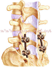
We plan each screw's position according to vertebral anatomy. Implant sizes, trajectories and construct length are selected for the particular procedure.
If treatment includes fusion, fixation supports the segment while bone fusion develops. Inserting metal instrumentation and subsequent fusion are different stages of treatment.[1]
When we consider spinal stabilisation
Fixation is discussed when support and stability are needed: for certain types of instability and vertebral slippage, deformities, fractures or reconstruction after decompression. The decision depends on diagnosis, symptoms, the condition of supporting structures and the planned operation.
Not everyone with lumbar stenosis needs additional stabilisation. We assess whether nerve decompression alone can preserve stability without instrumentation.[5]
Treatment of spondylolisthesis and vertebral slippage · Endoscopic treatment of lumbar stenosis
What is needed to plan fixation?
For preliminary review, send existing investigations and describe what interferes with movement, work or sleep. If a surgical plan has already been proposed, include it.
- MRI: assessment of neural structures, discs and areas of compression.
- CT: clarification of bony anatomy and planning implant insertion when needed.
- X-rays: spinal position and balance; dynamic radiographs when indicated.
- Records of previous surgery: the discharge summary, details of the implanted system and earlier scans.
If osteoporosis is suspected, we assess bone health separately: it affects screw purchase in bone and preparation for surgery.[6] We will determine the list of additional investigations after the consultation.
Send MRI/CT studies as complete DICOM series. How to prepare and send investigations to the professor.
How we insert percutaneous instrumentation
Through separate small access points, screw trajectories are prepared and screws are inserted into the planned vertebrae. Rods are then passed through and the connections secured. Implant position is checked with imaging during surgery.[4]
The endoscopic part of the procedure, if needed, addresses decompression and cage insertion. Screw fixation is performed percutaneously. At the consultation, I will explain exactly which stages your operation will involve.[3]
Before surgery, we discuss the expected benefits and risks, including those affecting neural structures, the wound and construct fixation. Anatomy, bone health and other medical conditions influence the plan.
Endoscopic cage insertion and fixation in one plan
The operation combines several related objectives: decompressing nerves, preparing the interbody space, placing a cage with bone graft material and holding the segment with screws and rods. For posterior interbody fusion, I insert the cage endoscopically. Percutaneous screw fixation supplements interbody support.[3]
For specific indications, I consider ALIF — anterior lumbar interbody fusion. This is a different approach for inserting an interbody implant; its selection is discussed according to anatomy and reconstruction goals.
More about endoscopic and minimally invasive stabilisation · Comparison of endoscopic and open approaches
How we choose an implant system
We select instrumentation according to anatomy, number of levels, loading and bone health. Patients should understand which vertebrae we plan to fix and what function each component serves.
From investigation review to surgery
- Send your existing records. Describe your symptoms and previous treatment. If MRI/CT has already been performed, include the complete DICOM series and report. You can start with a consultation even if you have not yet had imaging. Preliminary review of existing materials is free.
- Attend a consultation and examination. We clarify the need for fixation and other procedural stages.
- Agree on the extent of surgery. We determine the levels, approach and implants, and discuss expected changes in mobility.
- Obtain a preparation plan. Tests, assessment of other conditions, and anaesthetist and medication recommendations.
- Return to activity according to the plan. Follow-up appointments, gradually increasing activity and rehabilitation.
What we agree before cage insertion and fixation
Before surgery, we discuss which segments will be fixed, where the cage will be placed and the purpose of each component. We use this to clarify the organisational plan.
- Treatment location. Agree the facility, date and admission arrangements. Where consultations and surgery take place.
- Preparation and anaesthesia. Obtain your individual investigation list and the anaesthetist's recommendations.
- Cost estimate. Clarify whether surgery, anaesthesia, required implants and materials, hospital stay and follow-up visits are included.
- Discharge and further follow-up. Agree activity advice, the review date and a team contact for questions after treatment.
Recovery after pedicle screw fixation
Internal stability helps organise the start of movement after surgery. First getting up and walking are assisted by the team. Before discharge, you receive recommendations on everyday movements and further appointments.
- Walking and everyday tasks: gradually increasing activity according to how you feel.
- Office work and driving: an agreed return plan, breaks and consideration of medicines that affect reaction times.
- Physical work and sport: staged activity increases and assessment of readiness.
If the operation involves fusion, bone union develops over months. Early pain relief and readiness for full loading are assessed separately.[1] We determine discharge and return-to-work timings according to the extent of surgery and your condition.
Questions about cages and screw fixation
Do you always use a cage for endoscopic interbody fusion?
Yes. In my technique, a cage and bone graft material are essential components of interbody fusion. The cage provides support between vertebral bodies, while percutaneous screws and rods hold the segment.
Why is a cage important in disc degeneration?
When surgery involves disc removal and interbody fusion, the cage maintains the space between vertebrae and, with bone graft material, creates conditions for fusion. Degenerative MRI changes alone do not mean that this operation is needed: we assess symptoms, stability and the treatment goal.
Can screws be inserted through small incisions?
Yes, percutaneous fixation uses separate small access points. We assess its feasibility according to anatomy and the required extent of treatment.
Will spinal mobility remain?
Movement is limited in the fixed segments; in fusion surgery, they are intended to unite. The effect on everyday activity depends on the number of levels and the condition of the rest of the spine.
Will the metal instrumentation need removal later?
Removal is not an automatic next stage. The decision depends on the type of surgery, the condition of the instrumentation, healing and clinical indications.
Can I have MRI after fixation?
Yes. We use only implants that allow MRI under the manufacturer's conditions. Tell the MRI specialist about the instrumentation before the examination.[7]
Can I enquire after a previous operation?
Yes. Send current MRI/CT studies, the discharge summary and details of the previous instrumentation. At the consultation, we assess the cause of symptoms and possible next steps.
What determines the cost of fixation?
The number of levels, approach, implant system and combination with decompression or fusion. We clarify the plan and estimate after reviewing investigations and holding a consultation.
Discuss minimally invasive spinal stabilisation with the professor
Send Professor Iakiv Fishchenko your investigations and the proposed surgical plan, if you already have one. Preliminary review is free. At a consultation in Kyiv, we will discuss the role of a cage, the need for interbody fusion and options for endoscopic insertion with percutaneous fixation.
Send your scans to Professor Fishchenko
How to send MRI/CT studies · About the professor · My endoscopic stabilisation website
Book an in-person consultation is possible without an MRI already available. Start with a description of symptoms and existing records; we will determine any further investigations after assessment.
Medical sources and the professor's resources
- AAOS OrthoInfo: spinal fusion, implants and recovery.
- AAOS OrthoInfo: minimally invasive spine surgery.
- Professor Iakiv Fishchenko: endoscopy, ALIF and percutaneous stabilisation.
- AO Surgery Reference: percutaneous pedicle screw insertion.
- WFNS Spine Committee: indications for stabilisation in lumbar stenosis.
- CNS: osteoporosis assessment before spinal implant insertion.
- FDA: preparing for MRI with implants.
- AAOS OrthoInfo: PLIF/TLIF interbody fusion, cages and screw fixation.
- Professor Iakiv Fishchenko: the cage in his own endoscopic interbody fusion technique.