Published:
How the injury happened
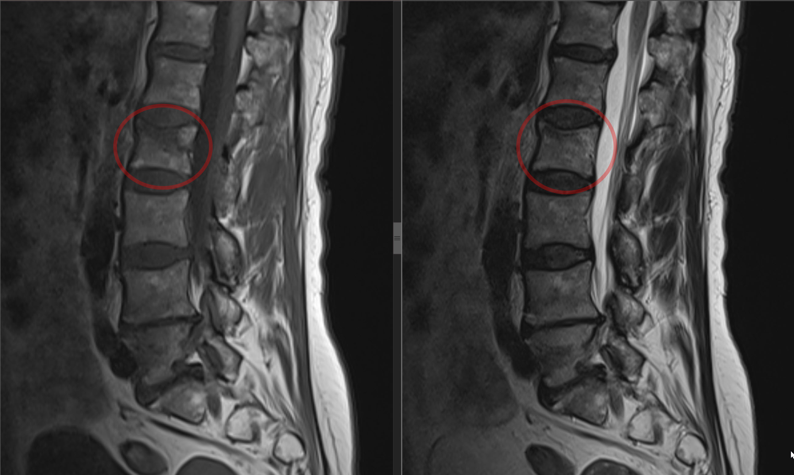
A 66-year-old woman fell onto her back when a bus braked suddenly. She developed severe lower back pain and rated it as 10 out of 10 when she moved. Imaging confirmed a recent compression fracture of the second lumbar vertebral body (L2).
Why I recommended vertebroplasty
The L2 vertebral body had lost little height, and there was no substantial deformity to correct. The main reason to intervene was the severe pain on movement. I recommended vertebroplasty to relieve that pain and help her get up and start walking sooner.
How the procedure was performed
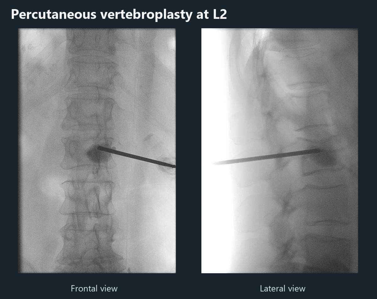
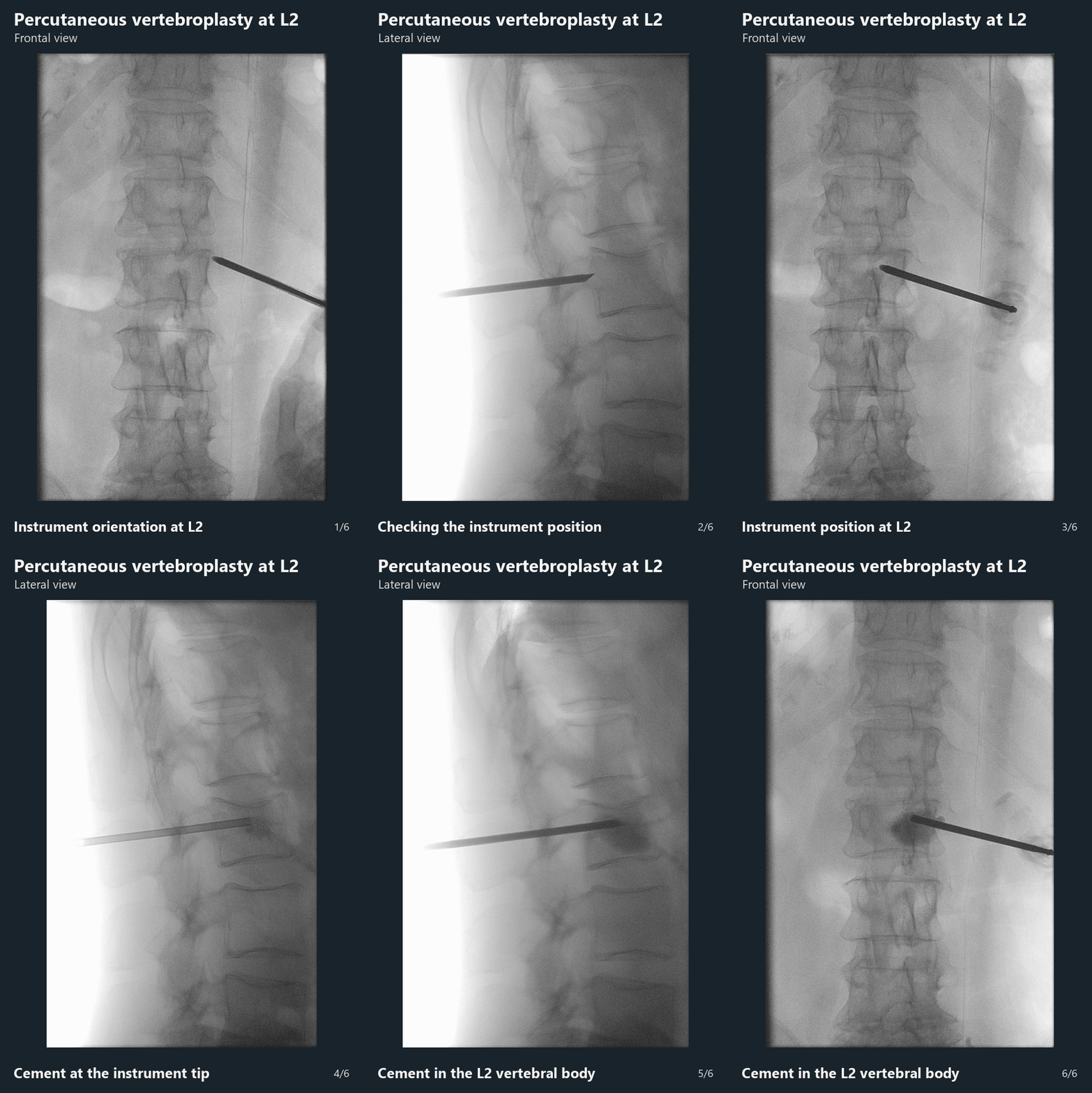
Using live X-ray guidance, I passed a needle through the pedicle on one side and injected 5 mL of bone cement directly into the L2 vertebral body. I did not use a balloon. The procedure took 15 minutes under propofol sedation.
What the follow-up images showed
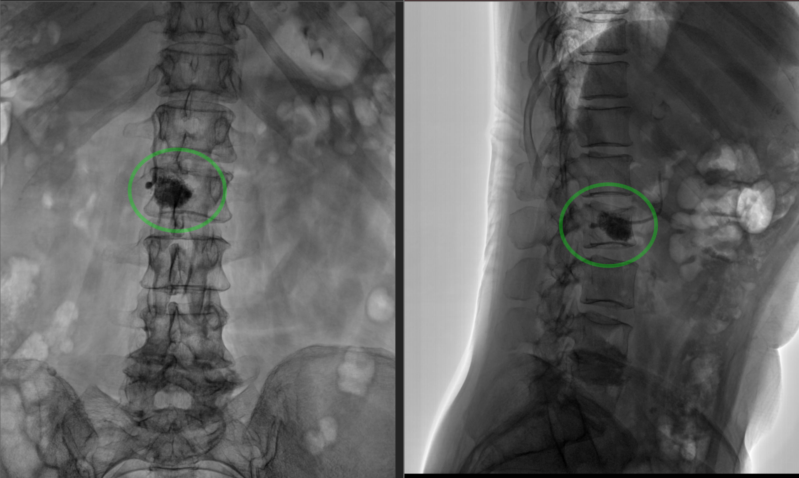
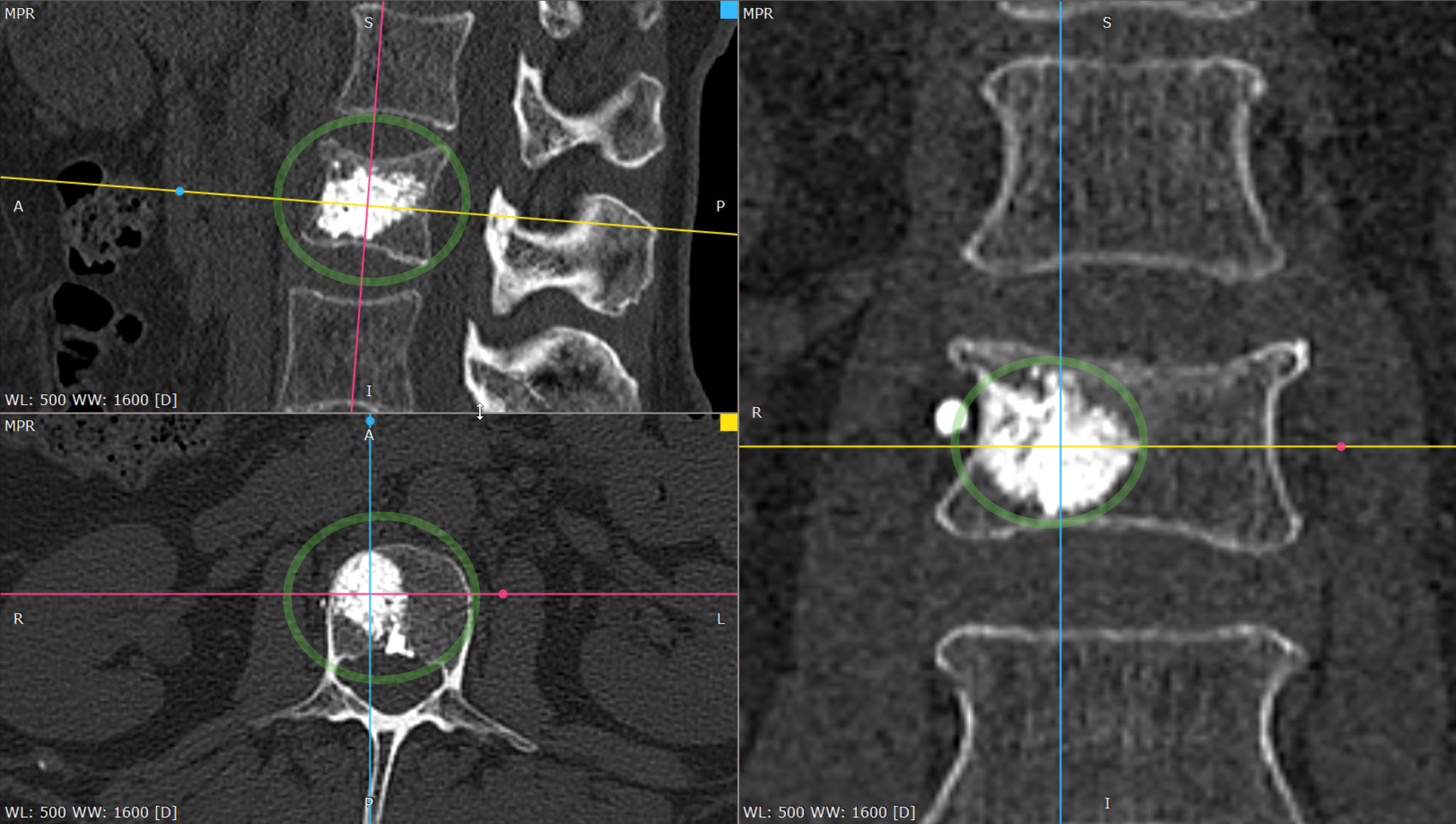
The postoperative X-rays showed the cement within L2. A CT scan the following day showed that some cement had entered a blood vessel. No cement entered the spinal canal, and the patient had no symptoms attributable to the vascular leakage. She was stable and able to stand independently at discharge from the spine unit. Further inpatient care with the thoracic surgery team was arranged for a separate injury to her diaphragm.
Pain and walking after the procedure
| Time | Clinical observation |
|---|---|
| Before the procedure | Lower back pain on movement was 10 out of 10. |
| One hour later | She took her first steps independently. |
| Within the first 24 hours | Lower back pain on movement was 3 out of 10. |
| At two weeks | She reported no lower back pain. |
The case description and images have been de-identified.
More about treatment
Discuss your case with the professor
Send a description of symptoms and existing investigations. If MRI/CT has already been performed, include the complete DICOM study and report. Preliminary review by the professor is free; an in-person consultation is also possible without an MRI already available.