
Does pain after a vertebral fracture make it difficult to get up, walk or sleep? Discuss percutaneous vertebroplasty in Kyiv with Professor Iakiv Fishchenko. The procedure aims to strengthen the painful damaged vertebra and help restore movement. Planning starts with image review and assessment of the cause of pain.
Vertebroplasty: strengthening a vertebra through a percutaneous approach
Percutaneous vertebroplasty is a minimally invasive procedure in which special bone cement is injected into the damaged vertebral body. Once hardened, it provides internal support at the fracture site. Access is through a small needle puncture, while instrument position and cement delivery are monitored with X-ray imaging.[1]
In my practice, compression fracture treatment includes vertebroplasty, balloon kyphoplasty and SpineJack. At consultation, I will explain which method fits your fracture anatomy and treatment aims.[4]
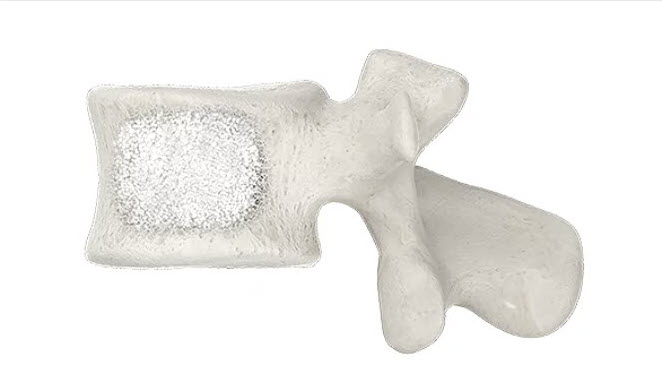
When we consider percutaneous vertebroplasty
Patients most often seek help for a painful osteoporotic compression fracture. When bone strength is reduced, a fracture can occur even after minor loading. It is important to establish that this particular vertebra is causing the pain.
- Pain significantly limits movement: getting out of bed, walking or performing usual activities is difficult.
- Prescribed pain relief does not help enough.
- Examination and images correspond to a painful fracture that has not yet healed.
In these circumstances, we discuss whether cement stabilization could help. A fracture on an image alone does not automatically require the procedure. Timing and extent are determined by symptoms, fracture age and general health.[2][3]
Which investigations to send to the professor
Start with the materials you already have. You do not need to arrange every investigation yourself before your first enquiry.
- CT or MRI in DICOM format: complete image series, the report and previous images if available.
- A description of symptoms: when and after what the pain began, where it is worst and whether you can get up and walk.
- Prescriptions and discharge summaries: what treatment you have received, which medicines you take and whether you have had spine surgery.
- Bone health information: results of osteoporosis investigations, if available.
CT helps assess bone structures and fracture shape; MRI helps assess fracture activity, bone marrow and neural structures. We will clarify the need for additional investigations after reviewing your materials.[3]
Step-by-step instructions: how to send CT/MRI scans in DICOM format
How vertebroplasty is performed
- Plan the procedure. We identify the painful level, approach and anaesthetic method.
- Administer anaesthesia. In my practice, vertebroplasty is performed under local anaesthesia with sedation. The anaesthetist agrees the final plan.
- Position the instrument. Through a percutaneous approach under X-ray guidance, we advance it into the vertebral body.
- Inject bone cement. We monitor its distribution at the fracture site; the cement gradually hardens.
- We monitor you after the procedure. We assess how you feel and whether you are ready to get up for the first time.
This is an image-guided percutaneous procedure. Its aim is fracture stabilization; separately identified neural compression or significant instability may require a different surgical plan.[1][4]
Vertebroplasty, kyphoplasty and SpineJack: what is the difference?
We choose the method according to vertebral condition, fracture age, deformity and required support. Height restoration and pain relief are different outcomes that we assess separately.[3][5]
Preparing for the procedure
After consultation, you receive an individual preparation plan. The team considers coexisting conditions, test results and medicines, particularly those affecting blood clotting.
- Tell us about regular medicines, allergies and previous reactions to anaesthesia.
- Agree with the anaesthetist when to stop eating and drinking.
- Change your medication only according to the agreed plan.
- Arrange an accompanying person and transport home after the procedure.
Additional tests and specialist consultations are determined according to your condition and the selected anaesthesia.[1]
Getting up for the first time and returning to activity
We help you get up for the first time after recovery from anaesthesia and assessment of how you feel, under the team's supervision. If your condition is stable, same-day discharge is possible; observation continues when needed.[4]
Before discharge, we agree on walking, everyday activities and follow-up. Return to work, exercises and lifting depends on the fracture, bone health and type of work. The need for a brace and its wearing schedule are determined individually.
The aim is to make movement more comfortable and gradually restore activity. Some patients experience rapid pain relief; we assess the pace of change at follow-up.[3]
Osteoporosis treatment after a fracture
Vertebroplasty strengthens one particular damaged vertebra. Preventing new fractures needs a separate bone-strength plan: osteoporosis assessment, appropriate treatment, safe activity and reducing fall risk.[3]
If you already take bone medication, send the prescription with your images. Information about osteoporosis.
What we discuss before choosing treatment
We assess expected pain relief, fracture anatomy and general health. Before the procedure, we explain risks including cement leakage outside the vertebra, bleeding, infection and injury to neural structures. X-ray guidance helps monitor instrument position and cement delivery.[1]
When urgent assessment is needed
If new or increasing leg weakness, urinary problems or loss of bowel control develops after injury or with a fracture, seek emergency medical help. Do not wait for a routine reply in a messenger.
Questions about percutaneous vertebroplasty
Can the original vertebral height be restored?
The main aim of vertebroplasty is to strengthen the fracture. If height correction is important, we separately assess kyphoplasty or SpineJack and what is possible for the specific fracture.
Can general anaesthesia be avoided?
In my practice, vertebroplasty is performed under local anaesthesia with sedation. The anaesthetist agrees the plan after assessing your condition.
Is an overnight hospital stay mandatory?
If your condition is stable after a check-up and getting up for the first time, same-day discharge is possible. The need for longer observation is determined individually.
What if I only have CT or only MRI?
Send what you already have as complete DICOM series, with the report and a description of symptoms. After review, I will explain whether additional investigations are needed.
What determines the cost of vertebroplasty?
The number of vertebrae, selected method, consumables, anaesthesia and observation required. We clarify the treatment plan and full cost after assessing investigations.
Discuss vertebroplasty with the professor
Send Professor Iakiv Fishchenko existing CT/MRI studies in DICOM format and a brief description of symptoms. State the injury date or when pain began, prescribed treatment and coexisting conditions. Preliminary review of your investigations is free.
Send CT/MRI scans to the professor
How to prepare investigations · About the professor · My compression fracture treatment website
Medical sources and the professor's resources
- ACR/RSNA RadiologyInfo: vertebroplasty and kyphoplasty.
- NICE TA279: selecting patients with painful osteoporotic fractures.
- NASS 2024: diagnosis and treatment of osteoporotic vertebral compression fractures.
- Professor Iakiv Fishchenko: treatment methods, anaesthesia and recovery.
- SAKOS: randomized comparison of expandable titanium implants and balloon kyphoplasty.