Published:
A clinical case from Professor Iakiv Fishchenko's practice
An 80-year-old woman. Leg pain and numbness limited walking to 10–15 metres. One month after endoscopic decompression, pain had decreased and walking distance was 150 metres.
The patient's presenting problem
According to the patient, low back pain had persisted for over a year and her condition was gradually worsening. The right leg hurt more, along the posterior and lateral thigh and in the groin. Left leg pain spread along its lateral aspect to the foot. Both legs were numb.
Conservative treatment did not provide lasting improvement.
Diagnosis and operation
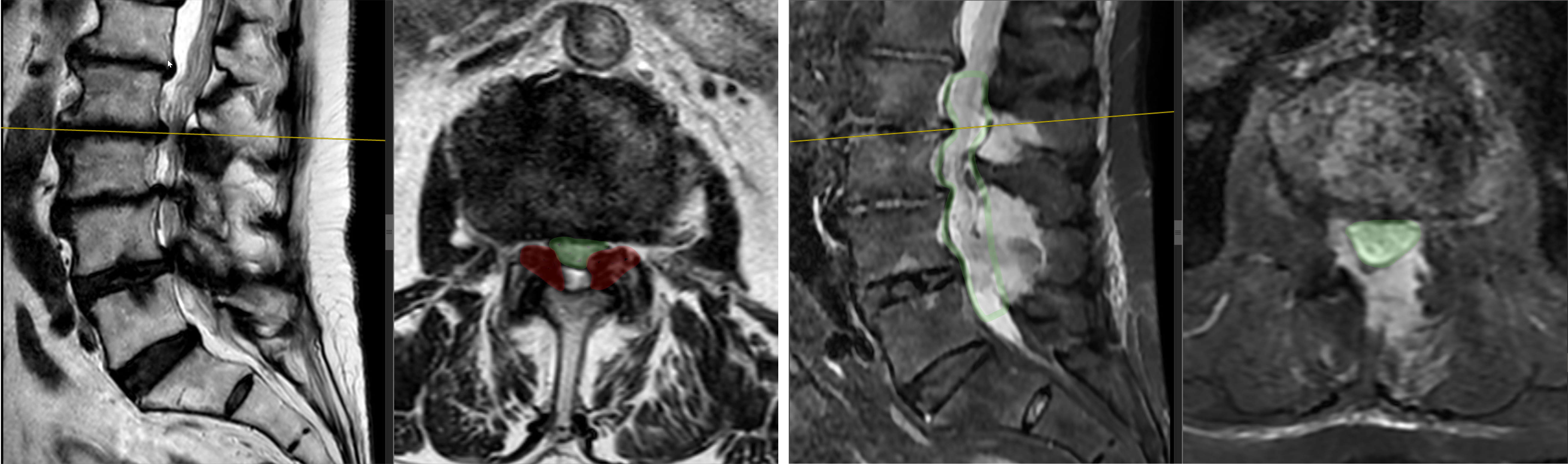
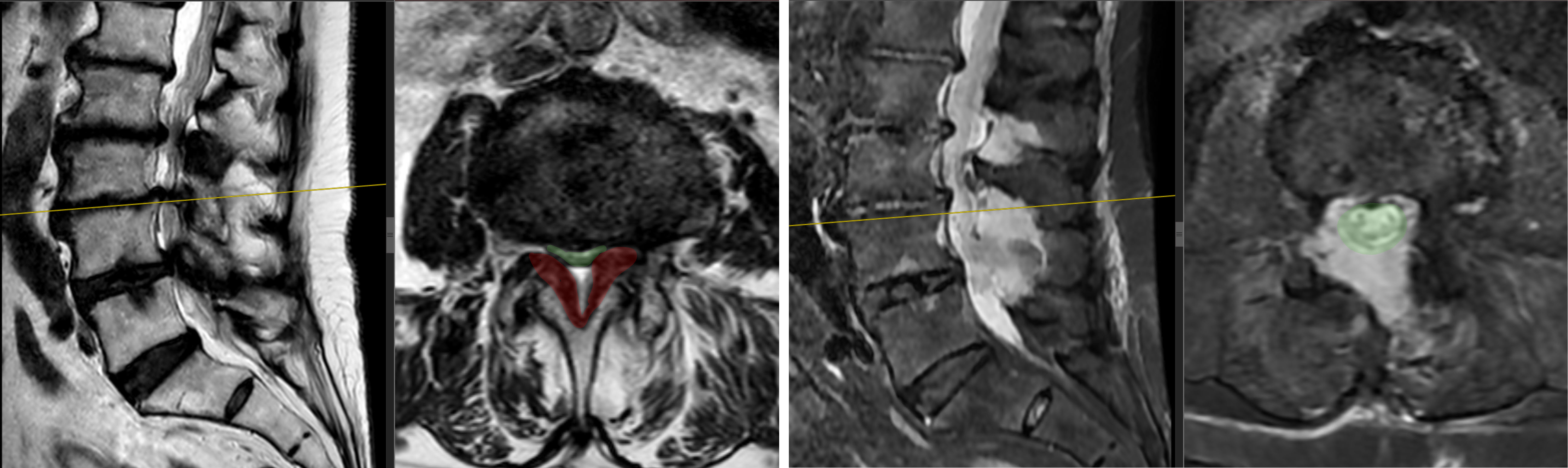
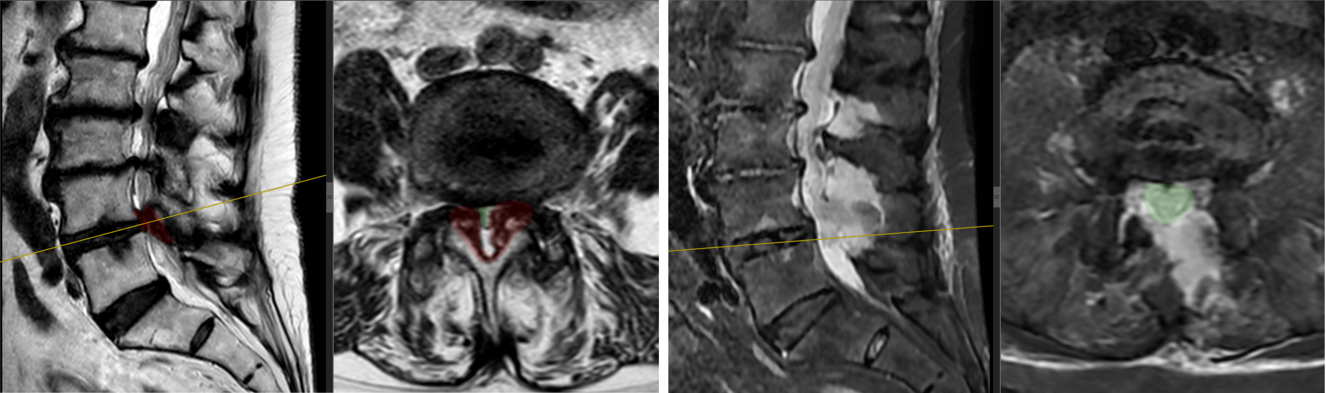
MRI showed spinal canal stenosis at L2–L3, L3–L4 and L4–L5. The diagnosis also included neurogenic claudication.
On 14 September 2026, L2–L3, L3–L4 and L4–L5 endoscopic biportal decompression was performed. Recovery was uneventful; the patient was discharged in satisfactory condition on the fourth day.
How she felt after one month
After one month, pain in the lower back and both legs had decreased. ODI and walking distance had also improved.
| Measure | Before surgery | After 1 month |
|---|---|---|
| Low back pain, VAS | 5–6 points | 2 points |
| Right leg pain, VAS | 8 points | 2 points |
| Left leg pain, VAS | 3–4 points | 1 point |
| ODI | 45% | 15% |
| Walking distance | 10–15 m | 150 m |
MRI before and after surgery
In each pair, preoperative images are on the left and postoperative images on the right. Slice angles and positions differ between the studies.
Click an image to open it at full size.
Clinical case author: Professor Iakiv Fishchenko.
Related information
Discuss your case with the professor
Send a description of symptoms and existing investigations. If MRI/CT has already been performed, include the complete DICOM study and report. Preliminary review by the professor is free; an in-person consultation is also possible without an MRI already available.