
Does your MRI report mention a vertebral haemangioma? At a consultation with Professor Iakiv Fishchenko in Kyiv, we will establish whether this is an incidental finding and whether further investigations or treatment are needed. If intervention is indicated, we will discuss strengthening the vertebra, decompressing the neural structures and planning a safe operation.
What is a vertebral haemangioma?
A haemangioma is a benign vascular formation within the bone of a vertebra. It is often discovered incidentally during MRI or CT performed for another reason.
The finding does not automatically identify the source of pain. At examination, we compare your symptoms with the scans and assess other possible causes: disc or joint changes, a fracture or nerve compression. The structure of the lesion, the condition of the vertebra and your symptoms matter in making a decision.[1]
Typical, atypical and aggressive haemangiomas
An atypical appearance alone does not establish aggressive behaviour. In a longitudinal MRI study, most haemangiomas grew slowly; decisions about an individual lesion require assessment of its images and the clinical situation.[2]
What MRI and CT show
MRI helps assess the structure of the lesion, soft tissues and its relationship to the nerves and spinal cord. CT shows the bony walls and internal vertebral architecture more clearly. Previous dated scans help assess changes.
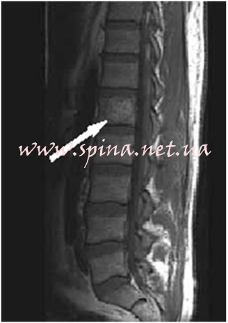
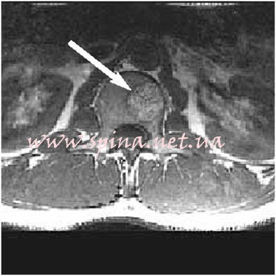
If features are uncertain, the doctor may arrange another imaging method, follow-up or consultation with a bone tumour specialist. The wording 'atypical haemangioma' alone does not determine management.[1]
When observation is sufficient
A confidently identified, typical, solitary incidental haemangioma in an adult without concerning features usually requires no treatment, additional investigations or regular follow-up MRI. This approach is included in the SSR Bone-RADS recommendations.[1]
If there is pain, uncertain structure or changes from previous scans, we clarify the plan individually. The aim of the consultation is to provide a clear answer: what needs attention and whether intervention is necessary.
When a haemangioma needs treatment
We discuss intervention when symptoms can reasonably be attributed to the lesion, vertebral support is damaged or neural structures are affected. We assess:
- persistent local pain with a cause consistent with examination and imaging findings;
- a fracture, destruction of vertebral walls or instability;
- extension of the lesion into the spinal canal and compression of nerves or the spinal cord;
- the onset or progression of neurological problems.
Diameter in millimetres or centimetres alone is not a universal indication for surgery. What matters is how the lesion changes the vertebra and whether it causes symptoms.[3][4]
New limb weakness, walking difficulties, saddle numbness or changes in bladder or bowel control require urgent medical assessment. Do not wait for a routine reply in a messaging app.
Vertebroplasty for a vertebral haemangioma
Percutaneous vertebroplasty involves injecting bone cement into the vertebral body through a small puncture under X-ray guidance. It may be used for selected painful haemangiomas to strengthen the vertebra and relieve symptoms.
Before the procedure, we assess the bony walls, the spinal canal and whether the pain is related to the lesion. If there is dangerous compression of neural structures, decompression and other treatment stages must be considered rather than limiting the plan to cement injection.[3]
At the consultation, I will explain what the procedure is intended to achieve and whether strengthening one vertebra is sufficient in your case.
How percutaneous vertebroplasty is performed · My specialist website about strengthening vertebrae
Decompression and stabilisation for an aggressive haemangioma
When the spinal cord or nerves are compressed, the aim of surgery is to decompress the neural structures. If the vertebra has lost its supporting function, reconstruction or fixation is needed. The extent of lesion removal and the combination of methods depend on its spread.
Because haemangiomas are vascular, bleeding control is planned in advance. In some cases, preoperative embolisation is considered to reduce the lesion's blood supply through the relevant vessels.[4]
A small approach must still allow the necessary nerve decompression and restoration of support. We decide on the approach and instrumentation after reviewing the complete scans and performing an in-person examination.
Embolisation, radiotherapy and other methods
Embolisation may be a stand-alone or preparatory stage of combined treatment. Its feasibility depends on vascular anatomy and the blood supply to the spinal cord.
Radiotherapy is considered for certain symptomatic or residual lesions together with a radiation oncologist. It does not replace urgent assessment when neurological problems are progressing.[5]
Sclerotherapy, including alcohol injection, has been described in specialist treatment series. This intervention has specific risks and requires selection and planning; it is not a universal injection for every haemangioma.[3]
The author's publication on procedural safety
In 2026, Audai Abudayeh and Iakiv Fishchenko published a scientific commentary in AME Medical Journal Vascularity before diagnosis in ambiguous compressive vertebral lesions. It addresses assessment of the vascularity of an indeterminate vertebral lesion before biopsy or decompression.
For patients, the practical message is this: the need for a biopsy and the access route are planned after assessing imaging and bleeding risk. A typical haemangioma often does not require biopsy. The publication is an expert letter to the editor.[6]
What to send for a consultation and how we plan follow-up
- Existing MRI/CT studies in DICOM format with the reports; previous dated scans, if available.
- Description of symptoms: where it hurts, how long it has been painful, what makes the pain worse, and whether there is numbness, weakness or a change in walking.
- Treatment history: previous procedures, discharge summaries and other important diagnoses.
After reviewing the records, I will explain whether further investigations, observation or surgery are needed. After an intervention, we agree on follow-up according to its type, symptoms and the condition of spinal support; there is no single schedule for everyone.
Step-by-step guide: how to prepare CT/MRI studies in DICOM format
Questions about spinal haemangiomas
Is a haemangioma cancer?
A haemangioma is a benign vascular lesion. If imaging does not allow this diagnosis to be established confidently, the nature of the finding needs clarification.
Does a haemangioma larger than 1 cm need surgery?
Size alone does not determine the indications. We assess vertebral structure, fractures, extension beyond the bone, neural structures and symptoms.
Must MRI be repeated every year?
For a confidently typical incidental finding without concerning features, regular MRI is often unnecessary. If the structure is uncertain or the clinical situation changes, follow-up is planned individually.
What does 'aggressive' mean in the report?
It describes the local behaviour of the lesion: expansion or destruction of bone, extension beyond it or an effect on neural structures. This calls for specialist assessment, rather than choosing a treatment yourself.
Find out whether your haemangioma needs treatment
Send Professor Iakiv Fishchenko your complete MRI/CT studies and a brief description of your symptoms. I will help assess whether the finding may explain your symptoms and determine further investigations and treatment options. Preliminary review of investigations is free.
Send your scans to Professor Fishchenko
How to prepare DICOM studies · About the professor · Contact and appointments in Kyiv
Medical sources and the author's publication
- SSR Bone-RADS, 2022: assessment of incidental solitary bone lesions in adults.
- Longitudinal MRI study of the natural history of vertebral haemangiomas.
- Primary study of combined treatment for symptomatic haemangiomas, 2023.
- Clinical series of surgical treatment for aggressive haemangiomas with neurological deficits.
- Radiotherapy for haemangiomas: a study of 80 patients, 2022.
- Abudayeh A, Fishchenko I. Vascularity before diagnosis in ambiguous compressive vertebral lesions. AME Medical Journal, 2026;11:31. Letter to the editor.