Patient: a 73-year-old woman. BMI: 28.0 kg/m².
Diagnosis: right-sided L5–S1 intervertebral disc herniation.
Operation: right-sided biportal endoscopic L5–S1 discectomy through an interlaminar approach.
Clinical findings
Pain down the back of the right leg had lasted more than six months. Before surgery, intensity was 8 on the visual analogue scale (VAS). Weakness of the right foot extensors was identified, with muscle strength graded 2/5 on the MRC scale.
The patient had completed two courses of physiotherapy and a course of non-steroidal anti-inflammatory drugs. She had not previously undergone lumbar spine surgery.
Preoperative imaging
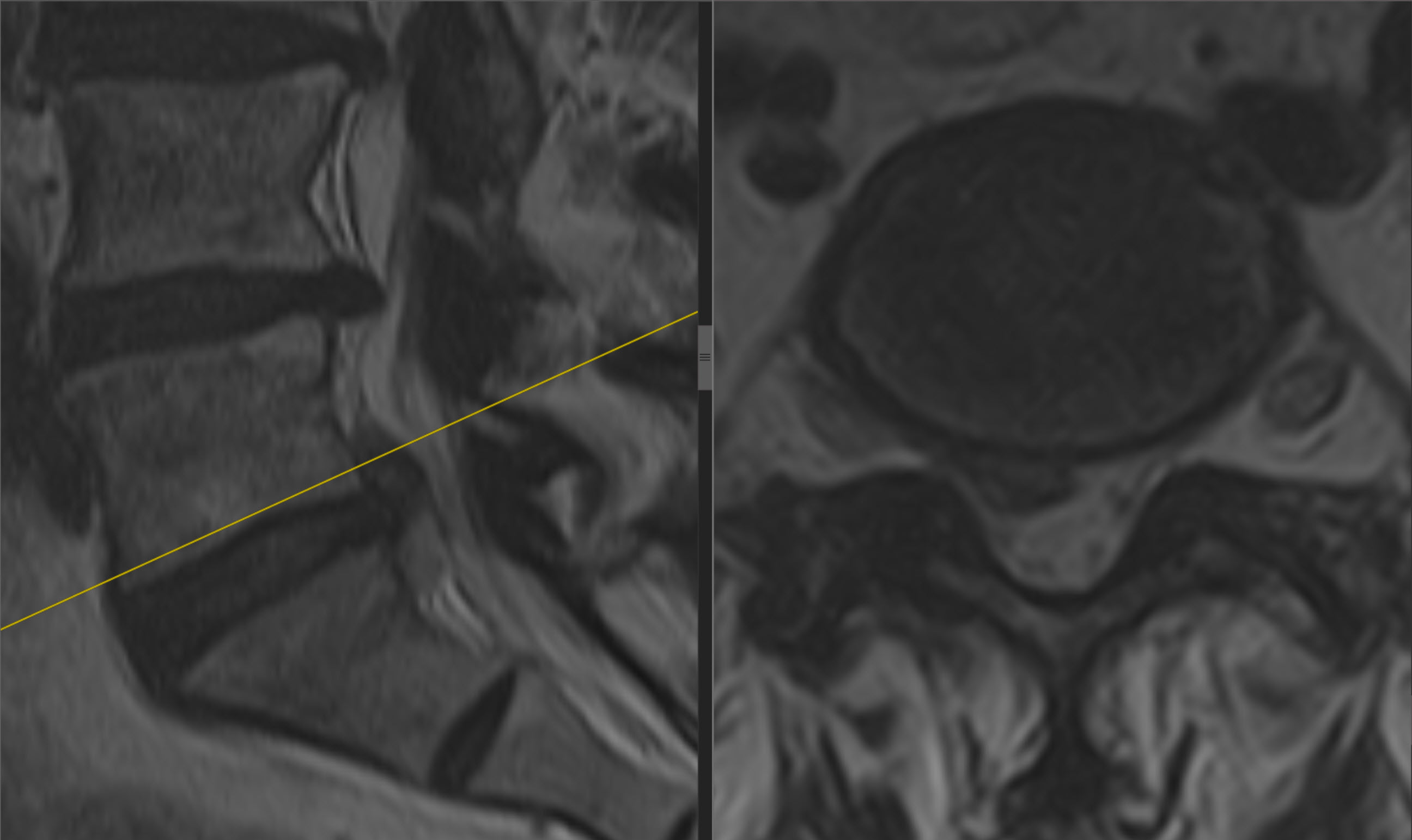
Surgical treatment
Right-sided biportal endoscopic L5–S1 discectomy was performed through an interlaminar approach. There were no intraoperative complications.
Postoperative course
The next day, right leg pain decreased to 5 on the VAS. Follow-up lumbar MRI was performed.
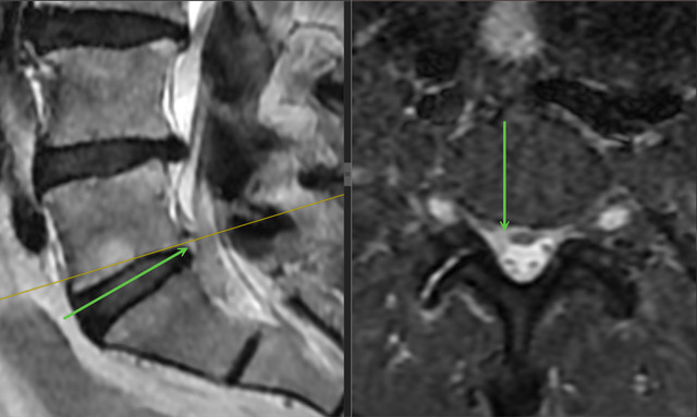
At one week, right leg pain had decreased to 2 on the VAS, and right foot extensor strength had increased to 3/5 on the MRC scale. There were no early postoperative complications.
Clinical case source
- The complete original case in the author's atlas, in Ukrainian
- The “Clinical examples” section of the author's atlas (original-language resource)
This describes one clinical case with its stated follow-up period. Its outcome is not a promise that another person's treatment will follow the same course.
Materials for contacting us
Discuss your case with the professor
Send a description of symptoms and existing investigations. If MRI/CT has already been performed, include the complete DICOM study and report. Preliminary review by the professor is free; an in-person consultation is also possible without an MRI already available.