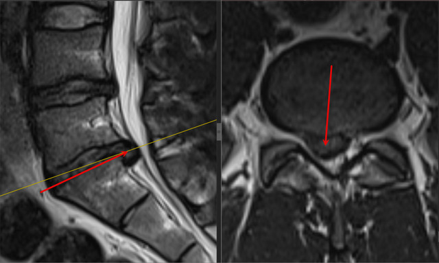
Patient: a 19-year-old man. BMI: 26.9 kg/m². Diagnosis: right-sided L5–S1 disc herniation. Operation: right-sided biportal endoscopic L5–S1 discectomy through an interlaminar approach.
Clinical findings
Right leg pain had lasted more than six months. Before surgery, intensity was 7–8 on the visual analogue scale (VAS). The right foot and toe extensors were weak, with strength graded 3/5 on the MRC scale. The patient had received conservative treatment. He had not previously undergone lumbar spine surgery.
Preoperative imaging
Surgical treatment
Right-sided biportal endoscopic L5–S1 discectomy using BESS/UBE was performed through an interlaminar approach. There were no intraoperative complications.
Postoperative course
The next day, right leg pain decreased to 3–4 on the VAS. Follow-up lumbar MRI was performed.
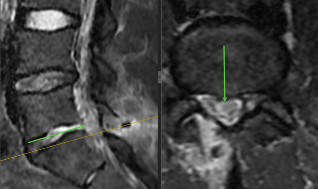
At one week, right leg pain had decreased to 2–3 on the VAS, and right foot and toe extensor strength was 5/5 on the MRC scale. There were no early postoperative complications.
Clinical case source
- The complete original case in the author's atlas, in Ukrainian
- The “Clinical examples” section of the author's atlas (original-language resource)
This describes one clinical case with its stated follow-up period. Its outcome is not a promise that another person's treatment will follow the same course.
Materials for contacting us
Discuss your case with the professor
Send a description of symptoms and existing investigations. If MRI/CT has already been performed, include the complete DICOM study and report. Preliminary review by the professor is free; an in-person consultation is also possible without an MRI already available.