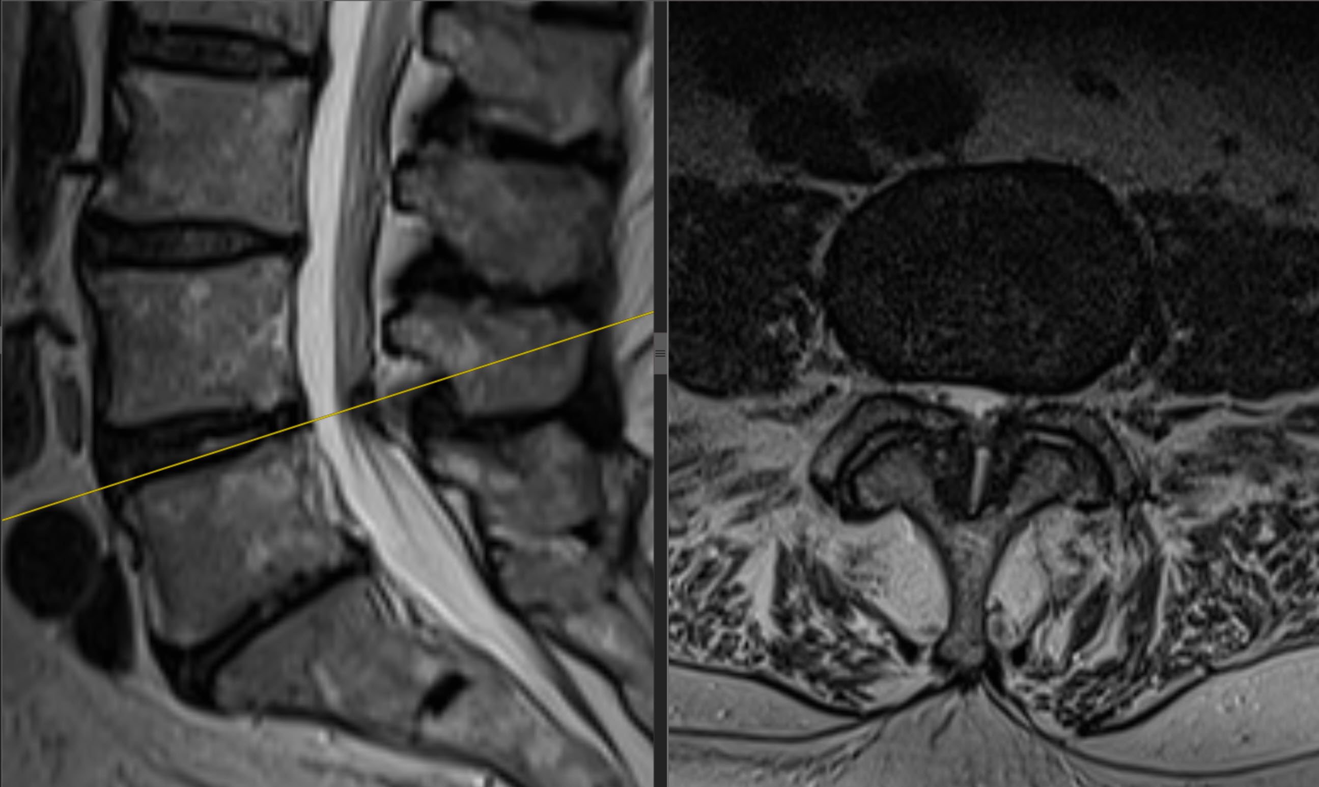
Patient: a 79-year-old woman. BMI: 42.2 kg/m². Diagnosis: absolute spinal canal stenosis at L4–L5.
Operation: biportal endoscopic L4–L5 decompression (BESS/UBE) from the left, without stabilisation.
Symptoms and history
For five months, lower back pain had spread into the legs when walking. Before surgery, intensity was 9 on the visual analogue scale (VAS), and walking distance was limited to approximately 20 metres. Urinary incontinence had also been present during the last two months before surgery.
There had been no previous lumbar spine operations.
Preoperative imaging
Surgical treatment
Biportal endoscopic L4–L5 decompression was performed using BESS/UBE through two separate portals from the left. The operation lasted 36 minutes. There were no intraoperative complications.
Stabilisation was not performed: the patient had no scoliotic deformity, spondylolisthesis or spinal instability. Age, osteoporosis and general condition were also considered when choosing the extent of surgery.
Watch the full operation recording on Vimeo
Video 1. Biportal endoscopic L4–L5 decompression without stabilisation. The first 30 seconds are an English introduction with clinical findings and pre- and postoperative MRI, followed by the full 36 min 29 sec operation recording. Total video duration: 36 min 59 sec.
Postoperative course
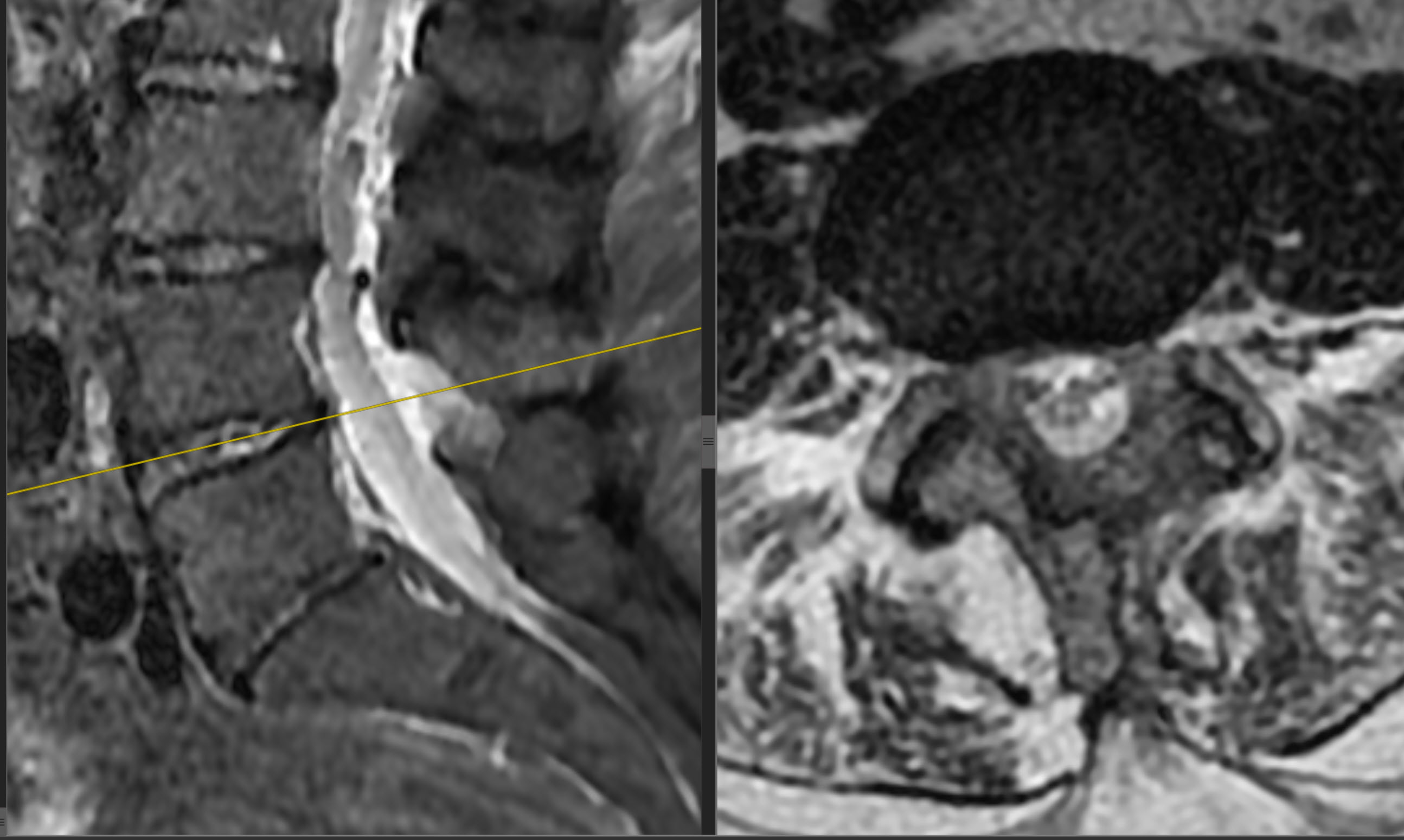
Follow-up MRI was performed the next day. In the surgeon's assessment, compression of the nerve structures at the operated level had been relieved.
On day three, pain had decreased to 3 on the VAS. After several days, the patient could walk 50 metres. The hospital stay lasted four days.
Urinary incontinence decreased substantially the next day and resolved completely within a week.
At one month, pain intensity was 2 on the VAS and walking distance was 200 metres. There were no postoperative complications. The latest available assessment was one month after surgery.
Author: Professor Iakiv Fishchenko.
Original source and case context
This describes one clinical case and the actual follow-up of this patient. The result is not a promise of the same course for someone else; treatment is planned individually.
Resources about this operation
Discuss your case with the professor
Send a description of symptoms, previous recommendations and existing investigations. If MRI/CT is available, send complete DICOM studies and reports. Preliminary review by the professor is free.
Send materials to the professor
How to prepare investigations in DICOM format · Book an in-person consultation in Kyiv