Does the back stay rounded even when the adolescent tries to straighten up? In Scheuermann's kyphosis, vertebral shape, remaining growth and progression of the curve need assessment. Professor Iakiv Fishchenko will help determine when observation or bracing is sufficient and when surgical correction should be discussed.
Scheuermann's kyphosis: a structural spinal deformity
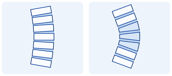
Scheuermann's disease is a structural increase in kyphosis, usually identified in adolescence. The front of several vertebral bodies becomes lower than the back: they become wedge-shaped and the spinal curve increases.
Ordinary slouching is more flexible and may improve when straightening up. In structural kyphosis, altered vertebral shape limits this correction. The cause of a rounded back cannot be determined from a photograph alone or explained solely by a habit of sitting poorly.[1]
Which changes need attention?
- A rounded or angular back that is difficult to straighten voluntarily.
- Back pain and fatigue, particularly after prolonged sitting, standing or exertion.
- A noticeable postural change during growth or an increase in the curve on successive images.
- Limited activity or dissatisfaction with appearance that affects everyday life.
Not everyone has pain. Treatment selection depends on the course of the deformity and its effect on life, rather than symptom severity on the day of consultation alone.[2]
When urgent assessment is needed
New leg weakness, walking problems, rapidly increasing numbness or new bladder or bowel control problems require urgent medical assessment. Do not wait for a routine reply in a messenger. These signs should not be regarded as ordinary slouching.
Diagnosis: examination and standing X-rays
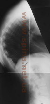
At your appointment, we assess posture, curve flexibility, trunk balance and neurological function. Standing whole-spine X-rays in frontal and lateral views help show the overall shape. The lateral image is used to measure kyphosis and assess vertebral body wedging.
Comparison with previous dated images helps determine whether the deformity is changing.[4] MRI and other investigations are ordered for a clinical purpose, particularly for neurological symptoms or surgical preparation.[1]
For preliminary review, send existing investigations. You do not need a new CT or MRI without a recommendation before your first enquiry.
When we discuss surgery for kyphosis
Surgical correction is considered for significant deformity, progression, persistent pain despite appropriate treatment or a substantial effect on function and quality of life. Neurological impairment requires separate assessment.
One X-ray angle does not determine the decision automatically. The location and rigidity of the curve, remaining growth, spinal balance, symptoms and expected benefit of surgery matter.[2]
At consultation, we will discuss what surgery can change, which levels need to be involved and how this will affect movement. The aim of planning is justified correction and preservation of function.
How surgical correction is performed
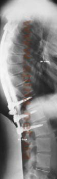
Correction of significant structural kyphosis usually combines curve correction, fixation and fusion. Screws and rods maintain the planned position, while bony union helps preserve correction. A posterior approach is most commonly used.
A rigid deformity may require osteotomies: controlled changes to bone structures that allow correction. Their necessity and the extent of fixation are determined by the individual anatomy.[4]
After fusion, movement decreases in the involved segments. Before treatment, expected changes in shape, trunk balance, recovery and risks are discussed, including infection, neurological injury or problems with fusion and the construct.[1]
How screws and rods work in spinal fixation · What fusion means
Observation, bracing and exercises
A brace is prescribed by a specialist; its schedule is adjusted at examinations. After growth is complete, a brace has less potential to change a structural deformity.[3] The exercise programme and permitted activity are selected according to symptoms and treatment aims.[5]
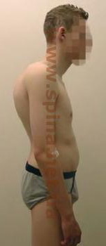
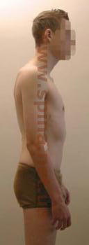
Archive photographs and X-rays
Materials from the previous version of the site illustrate the appearance of deformity and X-ray follow-up. They do not determine the method or expected extent of correction in your case.
What to send the professor for a consultation
- Existing standing X-rays of the entire spine in frontal and lateral views, with reports and dates. Include previous images for comparison.
- Age and a brief description of symptoms: when changes were noticed, whether growth is continuing, what limits activity and how posture is changing.
- Treatment history: bracing and exercise recommendations, previous examinations and operations. Send existing MRI/CT studies as complete series in DICOM format.
Preliminary review of the materials is free. They will help us determine the next step; a final plan requires clinical assessment and examination.
How to prepare and send existing CT/MRI studies in DICOM format · Consultation in Kyiv: contacts and booking
Recovery after surgery
Return to activity is planned according to the extent of correction, the patient's condition and the development of fusion. Pain control, gradual walking and examinations at specified times are important after surgery.
School, work, driving, sport and lifting are discussed separately. Each stage needs clear instructions; follow-up images help assess construct position and the course of recovery.[1]
Questions about Scheuermann's juvenile kyphosis
Is this caused by poor posture?
Scheuermann's disease changes vertebral structure. Poor posture may make slouching look worse, but does not fully explain the structural deformity.
Can kyphosis be corrected with exercises alone?
Exercises can improve comfort, strength and posture. The ability to affect a structural curve depends on its anatomy and growth; promising complete straightening through training alone would be inappropriate.
Is surgery necessary if the angle exceeds a certain number?
The decision considers the combination of angle and curve location, progression, pain, flexibility, age and impact on life. The angle is one part of assessment.
Can I seek advice as an adult?
Yes. We assess the current deformity, balance, symptoms and previous treatment. Persistent kyphosis alone does not make surgery mandatory.
Discuss a kyphosis treatment plan with the professor
Send Professor Iakiv Fishchenko your existing X-rays and a description of symptoms. We will review the possibilities of bracing, observation or surgical correction and determine the next steps. Preliminary review is free.
Medical sources
- AAOS OrthoInfo: types of kyphosis, investigations and treatment.
- POSNA: Scheuermann's kyphosis, its course and indications for treatment.
- AO Surgery Reference: exercises and bracing for Scheuermann's kyphosis.
- Scoliosis Research Society: Scheuermann's kyphosis, X-rays and surgical correction.
- NHS: kyphosis symptoms, physical activity and treatment options.