
Is it painful to get up, walk or turn in bed after a vertebral fracture? Discuss with Professor Iakiv Fishchenko in Kyiv how to strengthen the damaged vertebra and restore activity. Examination and imaging help us determine whether non-surgical treatment is sufficient or vertebroplasty, kyphoplasty, SpineJack or fixation is needed.
What is a spinal compression fracture?
In a compression fracture, the vertebral body is compressed and loses some of its height. One form is wedge deformity, in which the front collapses more. The degree of height loss matters, but choosing treatment also requires assessment of the posterior vertebral wall, ligaments, spinal canal and neural structures.
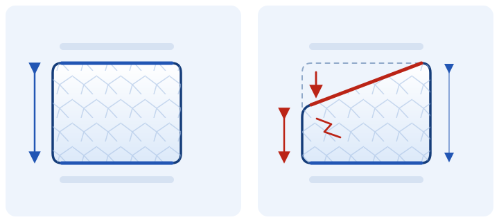
After major trauma, a fracture can occur even in bone of normal strength. In osteoporosis, it may follow a minor fall or everyday loading. If the findings are atypical, other causes of vertebral weakening, including tumour involvement, are assessed separately.[1]
Symptoms and situations that cannot wait
A fracture may cause new local back pain that worsens when getting up, standing or turning. Gradual loss of height or increasing stooping also warrants investigation. Sometimes a fracture is found on imaging without severe pain.
Emergency medical care is needed after serious trauma, or if there is new leg weakness, loss of sensation in the perineal area or urinary problems. Do not test your back with exercises, massage or forceful 'realignment'. Do not wait for a routine online reply.
If pain occurs alongside cancer, fever or an unexplained deterioration in general health, tell the doctor immediately.
CT and MRI: how we determine the type of fracture
Planning requires answers to three questions: which vertebra is damaged, whether stability is preserved and whether the current pain is caused by the fracture itself.
- Radiographs show vertebral height and shape and overall spinal alignment.
- CT shows the bony walls, fragments and fracture structure in greater detail.
- MRI helps assess bone marrow oedema, ligaments and neural structures. Oedema supports the assessment that the fracture is active, but does not establish the exact date of injury.
The need for each investigation depends on the situation. For your first enquiry, send what you already have: complete CT/MRI series in DICOM format, reports and previous scans, if available.[1]
When a vertebral fracture requires intervention
For a painful osteoporotic fracture, vertebral augmentation is considered if severe pain persists despite appropriate pain relief and examination and imaging confirm the painful level and an unhealed fracture. This corresponds to the NICE criteria for vertebroplasty and balloon kyphoplasty.[2]
After trauma, separate reasons for surgical assessment include instability, significant deformity and compression of neural structures. The decision cannot be reduced to a single percentage of height loss on an X-ray.[3]
There is no universal rule that everyone must wait the same number of weeks. The cause of the fracture, the course of the pain, ability to move and investigation results are important. At the consultation, I will explain the aim and extent of the proposed procedure.
Vertebroplasty, kyphoplasty and SpineJack
In my practice, these minimally invasive methods are used to strengthen appropriately selected fractures. Access is through punctures under X-ray guidance.
Strengthening the vertebra, correcting its height and relieving pain are different objectives. The SAKOS study compared SpineJack with balloon kyphoplasty in selected patients with osteoporotic fractures. Its findings do not establish one method for every type of injury.[4][5]
More about percutaneous vertebroplasty: the procedure, anaesthesia and recovery · My specialist website about treating vertebral fractures
When screws, rods and decompression are needed
An unstable injury requires restoration of spinal support. Pedicle screw and rod instrumentation may be used, including percutaneous insertion. If fragments or other structures compress the nerves, we plan their release — decompression — separately.
Cementing one vertebra does not address every aspect of a complex injury. Fixation levels and the need for additional reconstruction depend on injury morphology, ligament integrity and bone strength.[3]
When treatment without surgery is possible
Some stable fractures without neurological deficits are treated without surgery. The plan includes pain relief, agreed activity and follow-up. The need for a brace depends on the injury type and symptoms; it is not prescribed identically for everyone.[3]
The aim is a safe return to movement, rather than self-imposed prolonged bed rest. If pain worsens, deformity increases or new symptoms appear, reassessment is needed.
Returning to walking and everyday life
Once fracture stability has been established, we agree on first getting up, walking, everyday movements and gradually increasing activity. Exercises are selected according to the stage of recovery; for a painful osteoporotic fracture, early sessions may be performed in an unloaded position.[6]
Returning to work, driving and lifting depends on the fracture and treatment. Timings may differ after a percutaneous procedure and after fixation. At follow-up, we assess pain, mobility and, when needed, imaging.
How to reduce the risk of another fracture
If a vertebra fractured after minor loading, it is important to assess osteoporosis. Strengthening one vertebra does not treat reduced strength throughout the skeleton.
After a fracture, assessment of subsequent fracture risk, appropriate medication, falls prevention and safe activity are needed. Include any DXA bone density results and prescriptions in the records for your consultation.[6]
Questions about compression fractures
Does a compression fracture always need surgery?
No. Stability, neural structures and the cause of pain need assessment. Some fractures are treated without surgery; others require vertebral augmentation or fixation.
Can the vertebral height be restored completely?
The possibility of correction depends on the age and structure of the fracture. Vertebroplasty primarily strengthens the vertebra; we assess kyphoplasty and SpineJack separately for height correction.
Does an old fracture on MRI mean that it is the source of pain?
Not always. An old deformity may be an incidental finding. We compare the pain location, examination and signs of fracture activity, and assess other possible causes.
What should I do if I already have a fracture and the pain persists?
Send your existing CT/MRI studies in DICOM format, reports and a list of treatments. State when the pain began and whether you can get up and walk. After reviewing the records, I will explain the next step.
Discuss treatment of a vertebral fracture with the professor
Send Professor Iakiv Fishchenko your complete CT/MRI studies in DICOM format, reports and a brief description of your symptoms. State how the injury occurred or when the pain began, what treatment you have already received and whether osteoporosis is known. Preliminary review of investigations is free.
Send CT/MRI scans to the professor
How to prepare investigations · About the professor · Contact and appointments in Kyiv
Medical sources
- ACR Appropriateness Criteria: assessment and management of vertebral compression fractures.
- NICE TA279: indications for vertebroplasty and balloon kyphoplasty.
- WFNS Spine Committee: management of traumatic thoracolumbar fractures.
- ACR/RSNA RadiologyInfo: vertebroplasty and kyphoplasty.
- SAKOS: randomised comparison of SpineJack and balloon kyphoplasty.
- NOGG: management of symptomatic osteoporotic fractures, recovery and prevention of further fractures.