Patient: a 63-year-old woman. BMI: 35.2 kg/m².
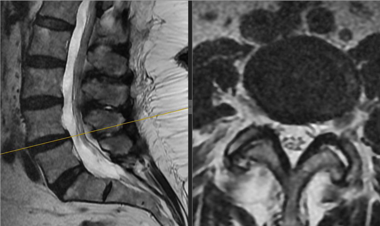
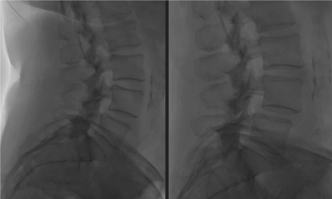
Diagnosis: L4–L5 spinal motion segment instability.
Symptoms and history
Severe lower back pain radiating into both legs for more than a year. Preoperative pain intensity: 7–9 on the VAS.
There was no leg muscle weakness. The patient did not report having to stop walking because of leg pain.
She had received repeated courses of conservative treatment from a neurologist and physical and rehabilitation medicine doctors, as well as epidural injections. Treatment provided minimal, short-lived relief. She had not previously undergone lumbar spine surgery.
Preoperative imaging
Lumbar MRI and flexion–extension X-rays.
Surgical treatment
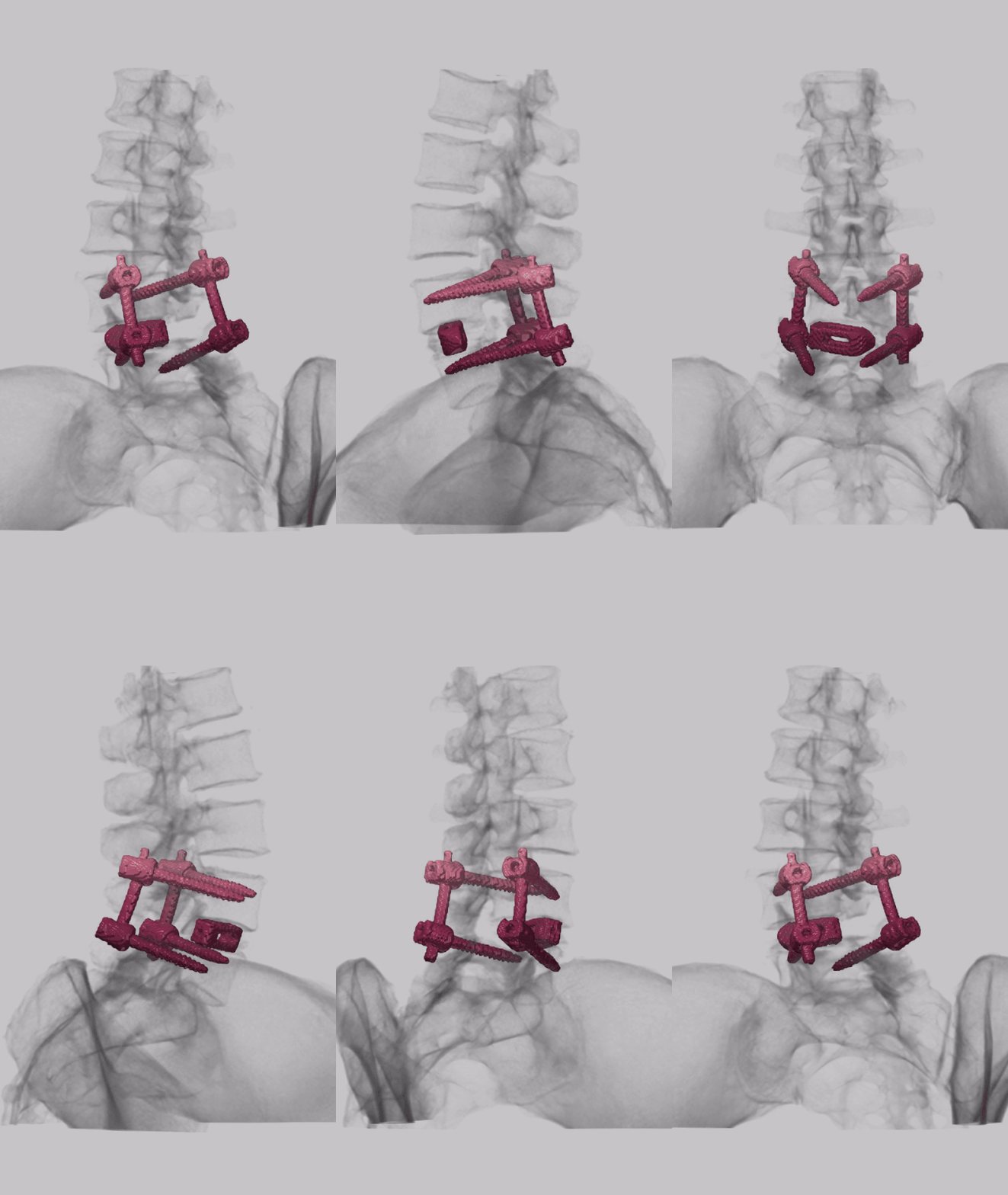
An L4–L5 interbody cage was inserted from the right side under endoscopic control, combined with percutaneous pedicle screw fixation.
There was no stenosis, so decompression was not performed.
Postoperative course
The next day, the patient stood and began walking. Lower back pain was 3–5 on the VAS, and leg pain was 3–4.
At one week, lower back pain was 3–4 and leg pain was 3 on the VAS.
There were no intraoperative or postoperative complications.
Postoperative imaging is presented as an animated three-dimensional CT reconstruction.
Show the animated CT reconstruction (20 MB)
Original source and case context
This describes one clinical case and the actual follow-up of this patient. The result is not a promise of the same course for someone else; treatment is planned individually.
Resources about this operation
Discuss your case with the professor
Send a description of symptoms, previous recommendations and existing investigations. If MRI/CT is available, send complete DICOM studies and reports. Preliminary review by the professor is free.
Send materials to the professor
How to prepare investigations in DICOM format · Book an in-person consultation in Kyiv