Published:
A clinical case from Professor Iakiv Fishchenko's practice
After disc removal, the patient developed acute right leg pain again. Revision surgery included endoscopic discectomy and L4–S1 stabilization.
The patient's presenting problem
The patient underwent L4–L5 discectomy in 2025. Symptoms returned in May 2026: pain radiated into the legs, worse on the right, along the lateral thigh and lower leg. Chronic low back pain had also persisted for several years, rated 4–6/10 on the VAS. Pain worsened at night, with bending and lifting. Conservative treatment did not help.
Diagnosis
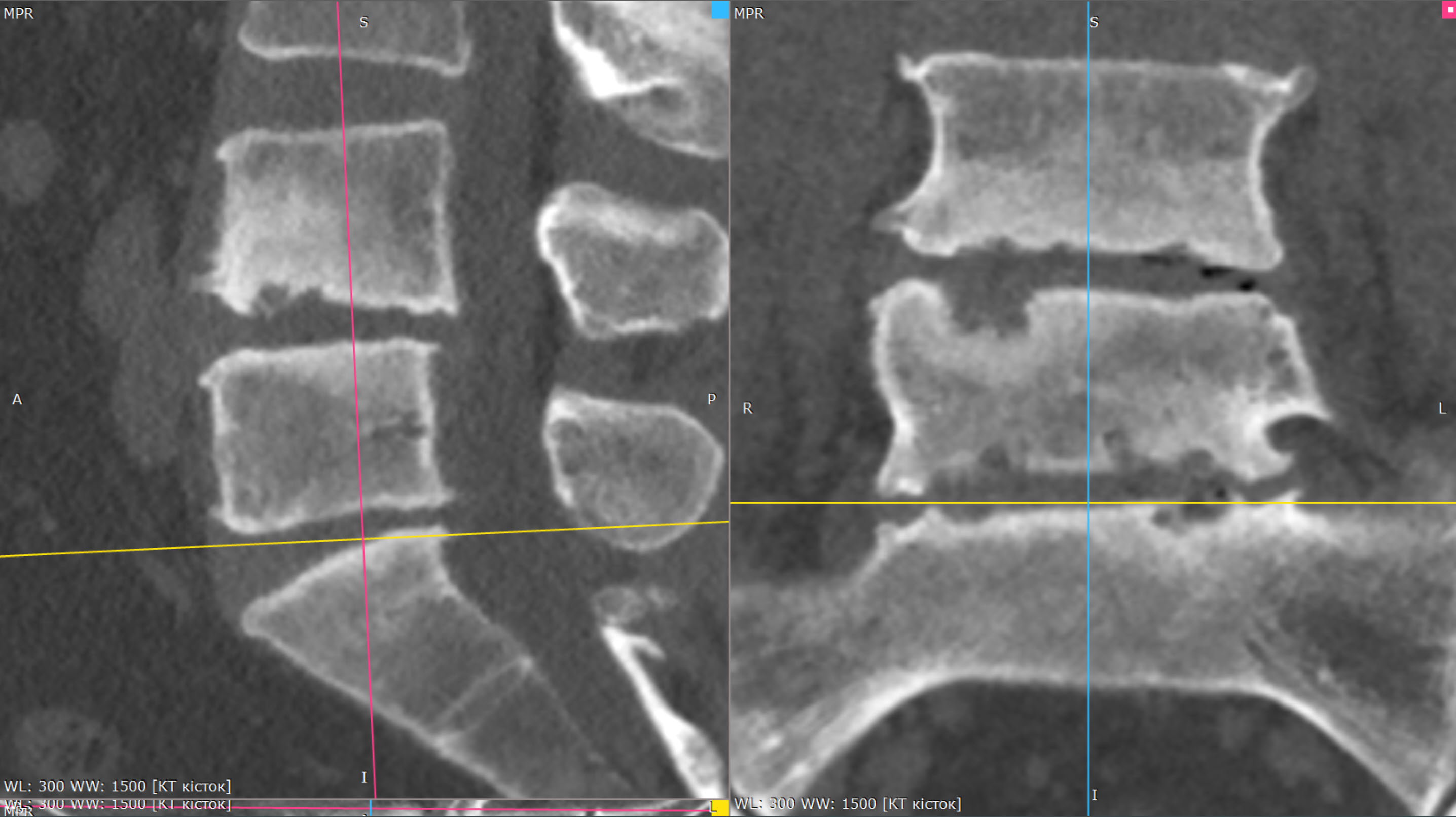
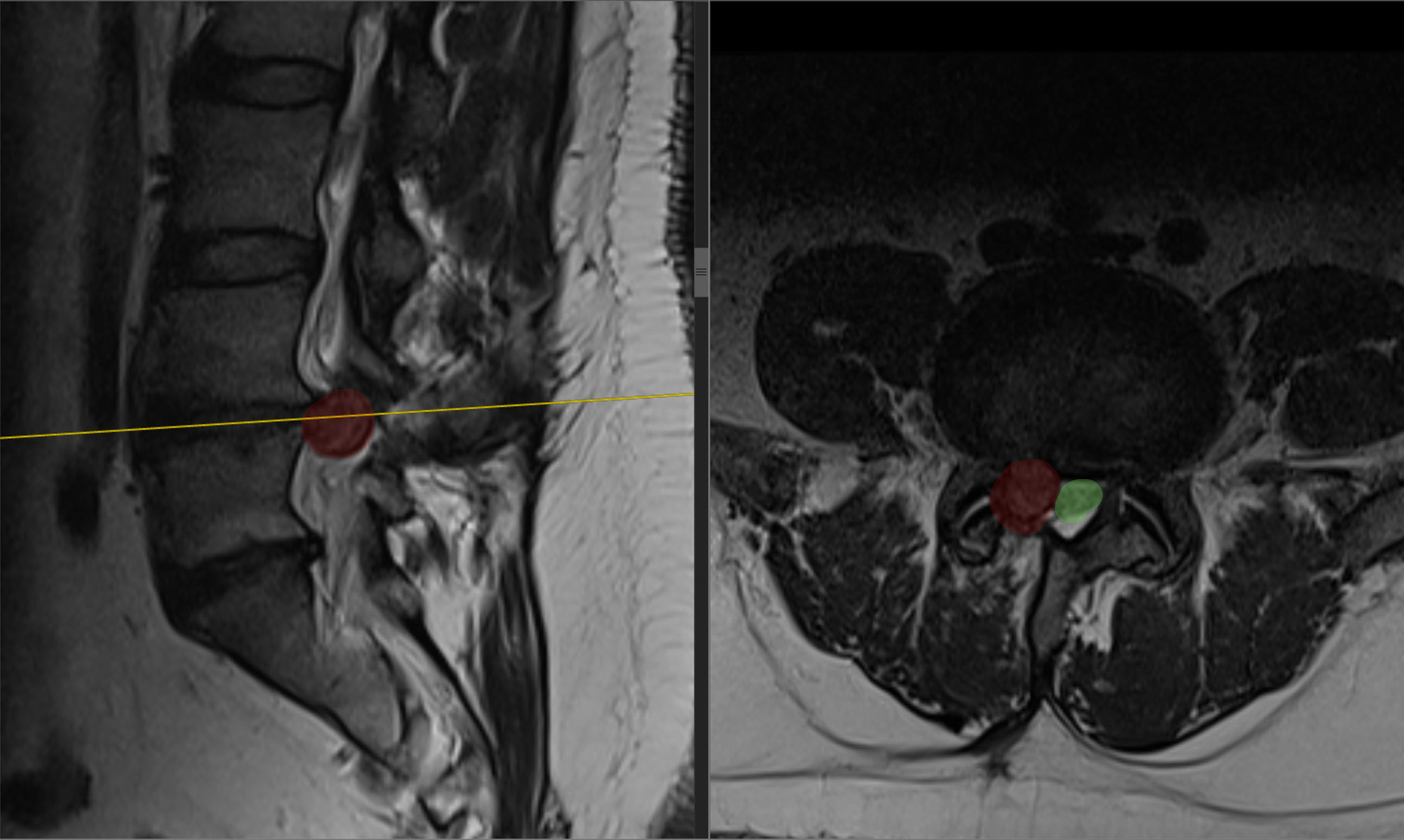
Recurrent L4–L5 disc herniation, marked degeneration of the L4–L5 and L5–S1 discs with endplate erosion, and L5 radiculopathy, worse on the right.
Choosing and performing surgery
Revision discectomy was supplemented with stabilization because recurrent L4–L5 herniation and marked degeneration of both discs with endplate erosion were accompanied by longstanding low back pain.
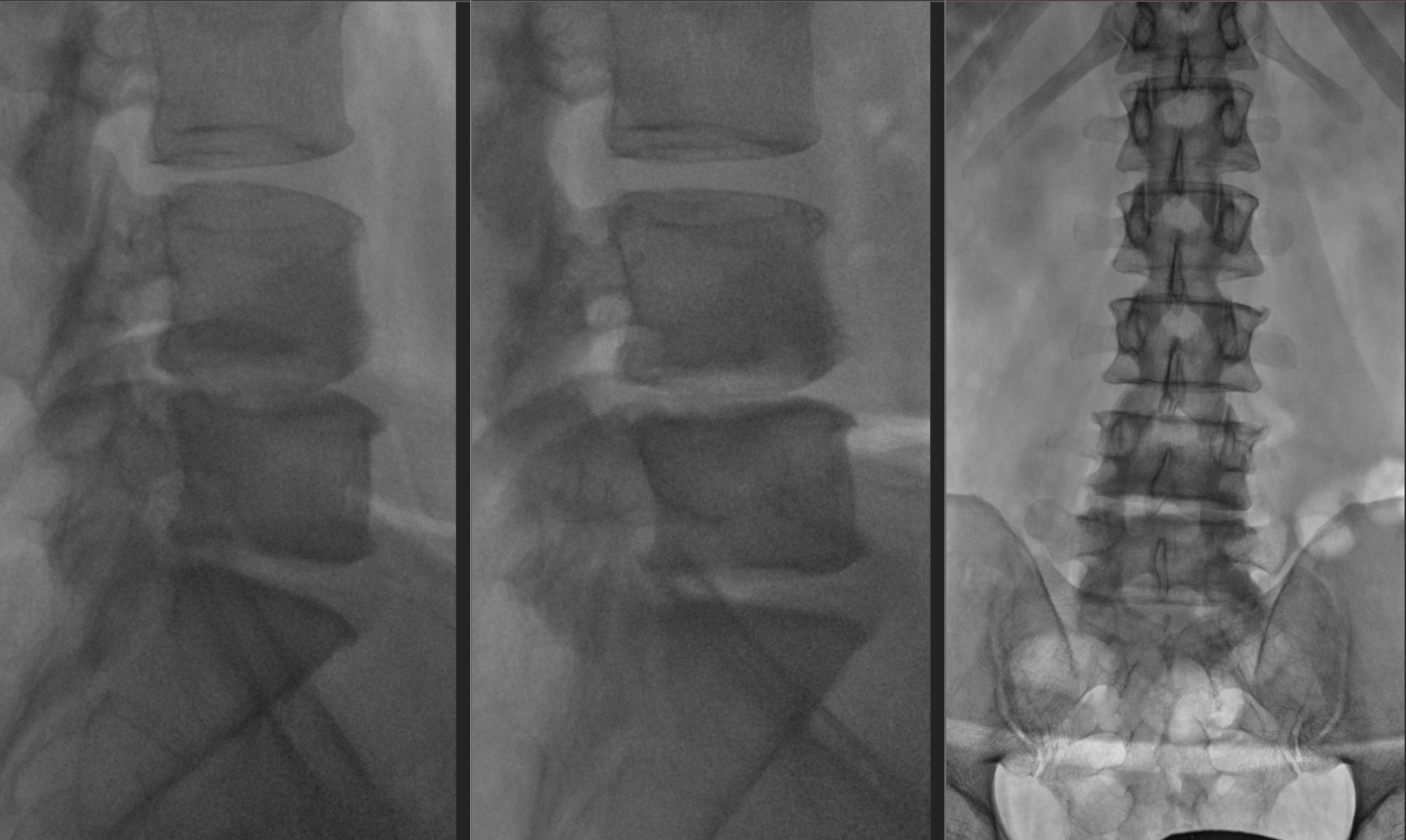
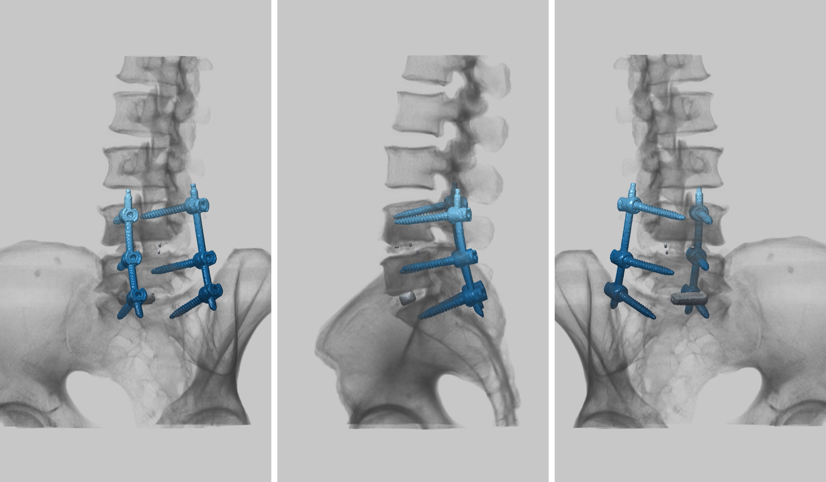
On 22 June 2026, L4–L5 and L5–S1 biportal endoscopic discectomy was performed at the Institute of Traumatology and Orthopaedics of the National Academy of Medical Sciences of Ukraine. It was combined with interbody fusion using MEDYSSEY cages (PEEK + titanium).
Posterior L4–L5–S1 fusion was also performed using a MEDYSSEY MIS pedicle screw construct.
MRI and X-rays
Outcome and postoperative CT
The postoperative course was uneventful. The wounds healed by primary intention. Seven days after surgery, on 29 June, the patient was discharged with improvement for further rehabilitation.
Clinical case author: Professor Iakiv Fishchenko.
Related information
Discuss your case with the professor
Send a description of symptoms and existing investigations. If MRI/CT has already been performed, include the complete DICOM study and report. Preliminary review by the professor is free; an in-person consultation is also possible without an MRI already available.