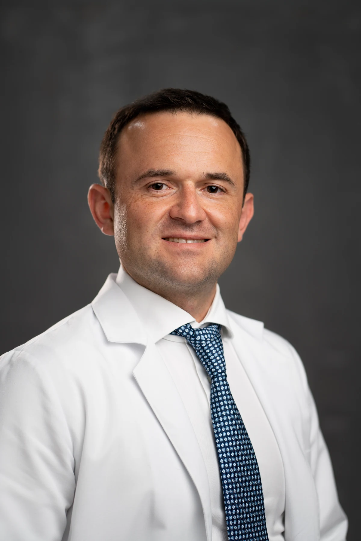
Lower back or leg pain, limited walking and trunk leaning can be symptoms of degenerative scoliosis. Professor Iakiv Fishchenko consults in Kyiv and assesses options for endoscopic nerve decompression, minimally invasive stabilisation and deformity correction. Start with a free preliminary review of existing investigations.
What is degenerative scoliosis?
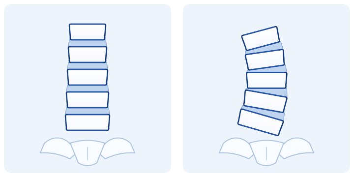
Degenerative, or de novo, scoliosis is spinal curvature that develops in adulthood because of uneven changes in the intervertebral discs and joints. It most commonly develops in the lumbar spine. Disc height decreases asymmetrically, vertebrae tilt and rotate, and load distribution changes.
We separately assess scoliosis that has existed since adolescence and later develops degenerative changes. Curve origin, mobility and trunk balance are important for planning.[1]
Symptoms: why the back hurts and walking becomes harder
Degenerative scoliosis may coexist with spinal canal stenosis and narrowing of the nerve root openings. At the consultation, we therefore assess both mechanical loading and nerve compression.[1]
- Lower back pain: worsens with standing, movement or exertion.
- Leg pain, numbness or weakness: may be related to nerve root compression.
- Reduced walking distance: you need to stop, sit down or bend forwards.
- A change in trunk position: leaning forwards or sideways, with difficulty standing upright.
Tell us what limits your life most: leg pain, back fatigue, walking or trunk position. This helps identify the main treatment goal.
Diagnosis and images for the consultation
Examination and existing investigations help match symptoms to specific spinal levels. Particularly important for deformity assessment are standing front and side X-rays of the entire spine, including the pelvis. They show the curve and body balance under load. MRI clarifies nerve compression, while CT assesses bony anatomy for specific indications.[1]
For initial contact, send what you already have:
- standing X-rays and previous dated images, if available;
- complete MRI/CT series in DICOM, together with reports;
- a description of pain, its duration and how far you can walk without stopping;
- information about previous treatment, surgery and implanted devices.
You do not need to arrange new investigations yourself before initial contact. After review, we will clarify which information is missing for planning.
Step-by-step instructions: how to send CT/MRI scans in DICOM format
When surgery is needed and how we choose its extent
We discuss surgery when symptoms substantially limit activity despite appropriate previous treatment, neurological deficit is progressing, or the deformity causes significant functional imbalance. One curve angle does not determine the decision.[2]
The extent of surgery must match the source of symptoms. Recommendations for decompression in ordinary stenosis cannot automatically be applied to every scoliotic curve.[2]
Endoscopic decompression for degenerative scoliosis
My specialisation is endoscopic spine surgery. The biportal technique uses a camera and instruments through separate small access points. A magnified image helps decompress nerve structures precisely while limiting exposure of surrounding tissues.
In stable degenerative scoliosis, treating stenosis is sometimes the main goal. In this situation, endoscopic decompression without major curve correction may be considered. It releases the nerves but does not by itself correct the entire deformity.
A 2026 retrospective study examined biportal decompression in 63 patients with stable degenerative scoliosis. It compared two intraoperative guidance methods; it does not prove that endoscopy replaces reconstruction in instability or substantial imbalance.[3]
Cages, screws and restoring spinal balance
If the plan includes interbody fusion, a cage supports the space between vertebrae and bone material provides conditions for fusion. Screws and rods hold the selected segments. These are distinct reconstruction components with a coordinated purpose.
In my endoscopic interbody fusion technique, the cage and bone material are essential. Posterior endoscopic cage placement can be combined with percutaneous fixation. With suitable anatomy, we separately consider ALIF: interbody implant placement through an anterior approach.
Reconstruction aims to provide support and the spinal alignment required. The approach, number of levels and need for additional decompression are determined during planning; using a cage does not automatically make an operation endoscopic.
Interbody cages · Pedicle screw fixation and an interbody cage · Treatment of vertebral slippage
When non-surgical treatment is an option
If there are no urgent indications, the plan may include selected activity, exercise and pain-control measures. Their aim is to maintain strength, endurance and daily activity. A brace is sometimes used for support in adults; it is not a guaranteed way to straighten an established curve.
Individual injection procedures are considered according to the pain source and expected benefit. They do not correct the deformity itself. If relief is insufficient, we reassess the cause of symptoms and whether surgery is appropriate.
Spinal injections and nerve blocks · Other forms of scoliosis and correction options
Preparation and recovery after surgery
Before fixation, bone condition needs assessment, particularly if there is suspected osteoporosis. This affects screw fixation and development of fusion. We determine the investigations and preparation needed from your information.[4]
We also consider general health, other medical conditions, medication and the extent of surgery needed. Before surgery, we agree expected benefits and risks, including infection, nerve injury and problems with fusion or the construct.[5]
The plan for returning to walking, work, driving and exertion depends on whether only decompression was performed or also fusion and correction. After reconstruction, recovery continues as healing and bony fusion develop. At follow-up visits, we assess function and adjust rehabilitation.
When emergency care is needed
New or rapidly increasing leg weakness, saddle numbness, urinary retention or loss of bladder or bowel control require emergency medical assessment. Do not wait for a routine reply in a messenger.
Questions about degenerative scoliosis
Does the entire curve always need correction?
No. If symptoms mainly arise from local stenosis and the deformity is stable, we consider a smaller procedure. With imbalance or instability, the surgical goals are broader.
Is MRI enough to choose an operation?
MRI shows nerve structures and surrounding tissues. Examination findings and standing X-rays are also needed to assess curvature and balance. Start by sending existing materials.
Can older people have surgery?
We plan according to symptoms, general health, bone strength and the extent of surgery needed. Age is considered alongside these factors, rather than as the only criterion.
Is dynamic ASC correction suitable?
ASC is not a universal method for every type of adult scoliosis. In degenerative deformity, we separately assess curve mobility, disc condition, stenosis and instability. These goals determine the correction option.
Discuss treatment for degenerative scoliosis
Send Professor Iakiv Fishchenko existing images, a description of pain and previous treatment. We will assess whether treatment can focus on endoscopic nerve decompression or also needs stabilisation or correction. Preliminary review is free.
Medical sources
- Scoliosis Research Society: adult scoliosis, symptoms and deformity assessment.
- WFNS Spine Committee: decompression, stabilisation and imbalance in lumbar stenosis.
- Liu et al., Orthopaedic Surgery, 2026: biportal decompression for stable degenerative scoliosis.
- Congress of Neurological Surgeons: preoperative osteoporosis assessment.
- Smith et al., Journal of Neurosurgery: Spine, 2021: five-year outcomes of surgical and non-surgical treatment of symptomatic adult lumbar scoliosis.
The professor's article on the Institute's website: endoscopic decompression, cage placement, ALIF and percutaneous fixation. It describes the techniques used in his practice; the spondylolisthesis studies cited are not separate outcomes of degenerative scoliosis treatment.