
Hand numbness, clumsy fingers or changes in walking may be signs of cervical stenosis. Professor Iakiv Fishchenko will compare your symptoms with MRI findings, explain the cause of compression and suggest a surgical treatment plan if needed.
What is cervical stenosis?
Cervical stenosis is narrowing of the space for the spinal cord or nerve roots. Causes can include disc changes, bone spurs and thickened ligaments. It is important to identify exactly where compression occurs and how it affects neural function.
These conditions may coexist. In myelopathy, attention to spinal cord function is more important than pain intensity alone.[1]
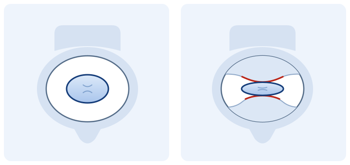
Symptoms of cervical myelopathy: pay attention to your hands and walking
Spinal cord compression can develop without severe neck pain. Reasons for an in-person assessment include:
- difficulty fastening buttons, writing or holding small objects;
- numbness or weakness in the arms or legs;
- unsteadiness, impaired balance or a change in your usual gait;
- gradual deterioration in everyday movements.
These symptoms can also have other causes. Examination is needed to establish their link to cervical compression.[1]
When emergency care is needed
Seek emergency medical help for rapidly increasing arm or leg weakness, sudden marked deterioration in walking, or new bladder or bowel problems. Do not wait for a reply in a messenger.[1]
MRI and examination: how we determine the cause of symptoms
At consultation, we assess strength, sensation, reflexes, hand movements and walking. Cervical MRI shows the spinal cord, discs and sites of compression. We compare the report with the actual images and examination.
When needed, CT clarifies bone changes; additional investigations are ordered according to the task. For preliminary review, send the materials you already have: you do not need to arrange the entire list of investigations yourself.[1]
When surgery is needed for cervical stenosis
The aim of decompression is to release the spinal cord or root from compression and preserve its function. For moderate and severe degenerative cervical myelopathy, AO Spine/CSRS guidelines recommend surgical treatment. There is no requirement to wait until every other method stops helping.
For mild myelopathy, surgery or structured rehabilitation with clinical monitoring is discussed. If neurological function deteriorates, the decision is reconsidered in favour of surgery. MRI compression without myelopathy or nerve root symptoms does not by itself mean preventive surgery is mandatory.[2]
At your appointment, I will explain what needs to be decompressed, how urgent treatment is and whether stabilization is also needed.
Operations for cervical stenosis: how the approach is chosen
We consider the location of compression, the number of affected levels, cervical alignment and vertebral stability. Several options are used in cervical stenosis surgery:
ACDF: anterior decompression and cage placement
The affected disc and structures compressing neural tissue are removed. A cage containing fusion material is placed between the vertebrae; fixation is selected to suit the construct. This combines decompression with fusion.
Corpectomy and reconstruction
When compression lies behind a vertebral body and access through the disc is insufficient, removal of part or all of the vertebral body may be necessary, followed by reconstruction of support and stabilization.
Posterior decompression: laminoplasty or decompression with fixation
These options enlarge the space for the spinal cord from behind, particularly in multilevel disease. Laminoplasty preserves some movement; when stabilization is required, decompression is combined with fixation and fusion.[3]
These are different ways of addressing a specific anatomical problem. We discuss an individual plan after analysing your images.
The role of endoscopy in cervical stenosis
My specialisation is endoscopic spine surgery. For suitable lateral or foraminal nerve root compression, endoscopic foraminotomy allows work through a small approach, releasing the nerve while preserving the natural disc.
The randomized study of posterior cervical endoscopy concerned lateral herniations with nerve root symptoms. Its results cannot automatically be applied to multilevel central spinal cord compression.[4]
In central stenosis, the priorities are adequate decompression and neck stability. ACDF or another selected reconstruction is planned around these aims. Endoscopy describes access and visualization; ACDF and disc replacement do not automatically become endoscopic procedures.
More about endoscopic cervical disc removal, ACDF and disc replacement · My cervical spine surgery website
What non-surgical treatment can offer
In selected cases with stable mild symptoms, symptom control, appropriate activity and structured rehabilitation with regular examination are possible. It is important to have a monitoring plan and know which changes require earlier assessment.[2]
Medicines or injections can affect pain, but do not widen a mechanically narrowed canal. If walking, strength or hand function deteriorate, do not replace spinal cord assessment with repeated courses of pain relief.[1]
What to send for a consultation with the professor
- Existing cervical MRI in DICOM format —all series and the report.
- A brief description of symptoms: what has changed in your hands, walking and balance, when it started and whether it is worsening.
- Other existing materials: CT, X-rays, previous examinations, postoperative discharge summaries and a medication list.
Preliminary review of investigations is free. An in-person neurological examination in Kyiv may be needed for a final plan.
Step-by-step instructions: how to send CT/MRI scans in DICOM format
What to expect from surgery and recovery
Decompression creates conditions for preserving and possibly improving function. Recovery depends on the condition of the spinal cord before surgery and the extent of the operation. We agree on walking, exercise, return to work and driving separately.[3]
Before surgery, we discuss expected changes, the effect of fusion on movement and the risks of the selected approach.
Questions about cervical stenosis
Is surgery necessary if the MRI report says “stenosis”?
The word alone is insufficient. We determine whether there is spinal cord or nerve root compression, clinical impairment and progression.
Can cervical myelopathy occur without severe pain?
Yes. Hand clumsiness or walking problems can be the main complaints even without marked neck pain.
Is a cage always used for stenosis?
A cage is part of ACDF with interbody fusion. Other decompression methods use different constructs; the need for support and fixation depends on the surgical plan.
Can a mobile disc replacement be used instead of ACDF?
In selected patients, yes. We assess movement, stability, joint condition and bone quality. Advanced degenerative changes may limit the possibility of disc replacement.[5]
Discuss a surgical plan for cervical stenosis
Send Professor Iakiv Fishchenko your existing MRI in DICOM format and a brief description of symptoms. We will review the sites of compression, the role of an endoscopic approach and the need for reconstruction or stabilization. Preliminary review is free.
Medical sources
- AAOS OrthoInfo: cervical myelopathy, symptoms and diagnosis.
- AO Spine/CSRS: guideline for the management of degenerative cervical myelopathy, 2017.
- AAOS OrthoInfo: surgery for cervical myelopathy and recovery.
- Ruetten et al., Spine, 2008: posterior cervical endoscopy for lateral disc herniations, randomized comparison.
- AAOS OrthoInfo: cervical disc replacement and patient selection.